{"title":"优化细胞迁移试验:荧光标记和趋化梯度的关键作用","authors":"Tomomi Tadokoro , Mimoko Kato , Tatsuya Kobayashi , Hideki Taniguchi","doi":"10.1016/j.bbrc.2024.150998","DOIUrl":null,"url":null,"abstract":"<div><div>Cell migration assays, also known as chemotaxis assays, are widely used to measure the migratory capacities of cancer cells, leukocytes, macrophages, and other motile cell types. In these assays, fluorescently labeled cells are seeded onto cell culture inserts with microporous membranes that block light transmission from 490 to 700 nm. The migrated cells are then observed and quantified from the bottom of the microporous membrane using a fluorescence microscope. In this study, we conducted cell migration assays using macrophages as the motile cells. We discovered that the commonly employed fluorescent labeling method using calcein acetoxymethyl ester (calcein AM) can lead to the time-dependent attenuation of fluorescent signals in certain cell types during migration assays, potentially compromising assay stability. This study overcame this limitation by utilizing PKH26, which fluorescently labels cell surfaces through a mechanism distinct from that of calcein AM. With this modification, we observed a consistent increase in the number of migrating macrophages over time. We also demonstrated that the gradient of chemoattractants is key to the success of cell migration assays. Our improved protocol provided reliable and stable results for cell migration assays.</div></div>","PeriodicalId":8779,"journal":{"name":"Biochemical and biophysical research communications","volume":"739 ","pages":"Article 150998"},"PeriodicalIF":2.5000,"publicationDate":"2024-11-14","publicationTypes":"Journal Article","fieldsOfStudy":null,"isOpenAccess":false,"openAccessPdf":"","citationCount":"0","resultStr":"{\"title\":\"Optimizing cell migration assays: Critical roles of fluorescent labeling and chemoattractant gradients\",\"authors\":\"Tomomi Tadokoro , Mimoko Kato , Tatsuya Kobayashi , Hideki Taniguchi\",\"doi\":\"10.1016/j.bbrc.2024.150998\",\"DOIUrl\":null,\"url\":null,\"abstract\":\"<div><div>Cell migration assays, also known as chemotaxis assays, are widely used to measure the migratory capacities of cancer cells, leukocytes, macrophages, and other motile cell types. In these assays, fluorescently labeled cells are seeded onto cell culture inserts with microporous membranes that block light transmission from 490 to 700 nm. The migrated cells are then observed and quantified from the bottom of the microporous membrane using a fluorescence microscope. In this study, we conducted cell migration assays using macrophages as the motile cells. We discovered that the commonly employed fluorescent labeling method using calcein acetoxymethyl ester (calcein AM) can lead to the time-dependent attenuation of fluorescent signals in certain cell types during migration assays, potentially compromising assay stability. This study overcame this limitation by utilizing PKH26, which fluorescently labels cell surfaces through a mechanism distinct from that of calcein AM. With this modification, we observed a consistent increase in the number of migrating macrophages over time. We also demonstrated that the gradient of chemoattractants is key to the success of cell migration assays. Our improved protocol provided reliable and stable results for cell migration assays.</div></div>\",\"PeriodicalId\":8779,\"journal\":{\"name\":\"Biochemical and biophysical research communications\",\"volume\":\"739 \",\"pages\":\"Article 150998\"},\"PeriodicalIF\":2.5000,\"publicationDate\":\"2024-11-14\",\"publicationTypes\":\"Journal Article\",\"fieldsOfStudy\":null,\"isOpenAccess\":false,\"openAccessPdf\":\"\",\"citationCount\":\"0\",\"resultStr\":null,\"platform\":\"Semanticscholar\",\"paperid\":null,\"PeriodicalName\":\"Biochemical and biophysical research communications\",\"FirstCategoryId\":\"99\",\"ListUrlMain\":\"https://www.sciencedirect.com/science/article/pii/S0006291X24015341\",\"RegionNum\":3,\"RegionCategory\":\"生物学\",\"ArticlePicture\":[],\"TitleCN\":null,\"AbstractTextCN\":null,\"PMCID\":null,\"EPubDate\":\"\",\"PubModel\":\"\",\"JCR\":\"Q3\",\"JCRName\":\"BIOCHEMISTRY & MOLECULAR BIOLOGY\",\"Score\":null,\"Total\":0}","platform":"Semanticscholar","paperid":null,"PeriodicalName":"Biochemical and biophysical research communications","FirstCategoryId":"99","ListUrlMain":"https://www.sciencedirect.com/science/article/pii/S0006291X24015341","RegionNum":3,"RegionCategory":"生物学","ArticlePicture":[],"TitleCN":null,"AbstractTextCN":null,"PMCID":null,"EPubDate":"","PubModel":"","JCR":"Q3","JCRName":"BIOCHEMISTRY & MOLECULAR BIOLOGY","Score":null,"Total":0}
Optimizing cell migration assays: Critical roles of fluorescent labeling and chemoattractant gradients
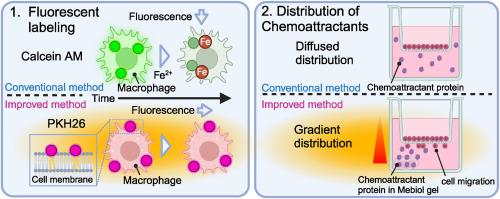
Cell migration assays, also known as chemotaxis assays, are widely used to measure the migratory capacities of cancer cells, leukocytes, macrophages, and other motile cell types. In these assays, fluorescently labeled cells are seeded onto cell culture inserts with microporous membranes that block light transmission from 490 to 700 nm. The migrated cells are then observed and quantified from the bottom of the microporous membrane using a fluorescence microscope. In this study, we conducted cell migration assays using macrophages as the motile cells. We discovered that the commonly employed fluorescent labeling method using calcein acetoxymethyl ester (calcein AM) can lead to the time-dependent attenuation of fluorescent signals in certain cell types during migration assays, potentially compromising assay stability. This study overcame this limitation by utilizing PKH26, which fluorescently labels cell surfaces through a mechanism distinct from that of calcein AM. With this modification, we observed a consistent increase in the number of migrating macrophages over time. We also demonstrated that the gradient of chemoattractants is key to the success of cell migration assays. Our improved protocol provided reliable and stable results for cell migration assays.
期刊介绍:
Biochemical and Biophysical Research Communications is the premier international journal devoted to the very rapid dissemination of timely and significant experimental results in diverse fields of biological research. The development of the "Breakthroughs and Views" section brings the minireview format to the journal, and issues often contain collections of special interest manuscripts. BBRC is published weekly (52 issues/year).Research Areas now include: Biochemistry; biophysics; cell biology; developmental biology; immunology
; molecular biology; neurobiology; plant biology and proteomics