Changrui Su, Wenlong Tang, Jinsheng Qiao, Wenchao Liu, Bin Hu, Kangda Huang, Qingguo Liu, Long Wang
{"title":"内窥镜经鼻腔经眶底至眶下区的泪囊前凹入路:解剖学研究","authors":"Changrui Su, Wenlong Tang, Jinsheng Qiao, Wenchao Liu, Bin Hu, Kangda Huang, Qingguo Liu, Long Wang","doi":"10.1007/s00405-024-08932-2","DOIUrl":null,"url":null,"abstract":"<h3 data-test=\"abstract-sub-heading\">Purpose</h3><p>The aim of this study is to describe the maximum exposure of the infraorbital region via the orbital floor using the transnasal prelacrimal recess approach (PLRA), and to provide an anatomical basis for treating lesions in the infraorbital region.</p><h3 data-test=\"abstract-sub-heading\">Methods</h3><p>Ten freshly injected frozen heads were dissected using the PLRA. The orbital floor was removed along the border of the medial infraorbital quadrangle, and the periorbita was opened to expose the infraorbital region. The areas of the medial infraorbital quadrangles were measured and analyzed. The PLRA was applied separately on the left and right sides of each cadaver head, resulting in a total of 20 prelacrimal recess approaches.</p><h3 data-test=\"abstract-sub-heading\">Results</h3><p>The PLRA enabled visualization of the optic nerve and the central retinal artery through the orbital floor. By integrating both medial and lateral approaches in relation to the inferior rectus muscle, all crucial anatomical structures within the infraorbital region could be clearly identified. The area of the medial infraorbital quadrangle was 420.65 ± 24.03 mm<sup>2</sup>.</p><h3 data-test=\"abstract-sub-heading\">Conclusion</h3><p>The PLRA provides access through the orbital floor to the maximum boundary of the infraorbital region, including the lateral orbital wall at the outermost level, the superior rectus muscle at the topmost level, and the medial orbital wall at the innermost level.</p>","PeriodicalId":11952,"journal":{"name":"European Archives of Oto-Rhino-Laryngology","volume":null,"pages":null},"PeriodicalIF":1.9000,"publicationDate":"2024-09-16","publicationTypes":"Journal Article","fieldsOfStudy":null,"isOpenAccess":false,"openAccessPdf":"","citationCount":"0","resultStr":"{\"title\":\"Endoscopic transnasal prelacrimal recess approach via the orbital floor to the infraorbital region: an anatomical study\",\"authors\":\"Changrui Su, Wenlong Tang, Jinsheng Qiao, Wenchao Liu, Bin Hu, Kangda Huang, Qingguo Liu, Long Wang\",\"doi\":\"10.1007/s00405-024-08932-2\",\"DOIUrl\":null,\"url\":null,\"abstract\":\"<h3 data-test=\\\"abstract-sub-heading\\\">Purpose</h3><p>The aim of this study is to describe the maximum exposure of the infraorbital region via the orbital floor using the transnasal prelacrimal recess approach (PLRA), and to provide an anatomical basis for treating lesions in the infraorbital region.</p><h3 data-test=\\\"abstract-sub-heading\\\">Methods</h3><p>Ten freshly injected frozen heads were dissected using the PLRA. The orbital floor was removed along the border of the medial infraorbital quadrangle, and the periorbita was opened to expose the infraorbital region. The areas of the medial infraorbital quadrangles were measured and analyzed. The PLRA was applied separately on the left and right sides of each cadaver head, resulting in a total of 20 prelacrimal recess approaches.</p><h3 data-test=\\\"abstract-sub-heading\\\">Results</h3><p>The PLRA enabled visualization of the optic nerve and the central retinal artery through the orbital floor. By integrating both medial and lateral approaches in relation to the inferior rectus muscle, all crucial anatomical structures within the infraorbital region could be clearly identified. The area of the medial infraorbital quadrangle was 420.65 ± 24.03 mm<sup>2</sup>.</p><h3 data-test=\\\"abstract-sub-heading\\\">Conclusion</h3><p>The PLRA provides access through the orbital floor to the maximum boundary of the infraorbital region, including the lateral orbital wall at the outermost level, the superior rectus muscle at the topmost level, and the medial orbital wall at the innermost level.</p>\",\"PeriodicalId\":11952,\"journal\":{\"name\":\"European Archives of Oto-Rhino-Laryngology\",\"volume\":null,\"pages\":null},\"PeriodicalIF\":1.9000,\"publicationDate\":\"2024-09-16\",\"publicationTypes\":\"Journal Article\",\"fieldsOfStudy\":null,\"isOpenAccess\":false,\"openAccessPdf\":\"\",\"citationCount\":\"0\",\"resultStr\":null,\"platform\":\"Semanticscholar\",\"paperid\":null,\"PeriodicalName\":\"European Archives of Oto-Rhino-Laryngology\",\"FirstCategoryId\":\"3\",\"ListUrlMain\":\"https://doi.org/10.1007/s00405-024-08932-2\",\"RegionNum\":3,\"RegionCategory\":\"医学\",\"ArticlePicture\":[],\"TitleCN\":null,\"AbstractTextCN\":null,\"PMCID\":null,\"EPubDate\":\"\",\"PubModel\":\"\",\"JCR\":\"Q2\",\"JCRName\":\"OTORHINOLARYNGOLOGY\",\"Score\":null,\"Total\":0}","platform":"Semanticscholar","paperid":null,"PeriodicalName":"European Archives of Oto-Rhino-Laryngology","FirstCategoryId":"3","ListUrlMain":"https://doi.org/10.1007/s00405-024-08932-2","RegionNum":3,"RegionCategory":"医学","ArticlePicture":[],"TitleCN":null,"AbstractTextCN":null,"PMCID":null,"EPubDate":"","PubModel":"","JCR":"Q2","JCRName":"OTORHINOLARYNGOLOGY","Score":null,"Total":0}
Endoscopic transnasal prelacrimal recess approach via the orbital floor to the infraorbital region: an anatomical study
Purpose
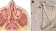
The aim of this study is to describe the maximum exposure of the infraorbital region via the orbital floor using the transnasal prelacrimal recess approach (PLRA), and to provide an anatomical basis for treating lesions in the infraorbital region.
Methods
Ten freshly injected frozen heads were dissected using the PLRA. The orbital floor was removed along the border of the medial infraorbital quadrangle, and the periorbita was opened to expose the infraorbital region. The areas of the medial infraorbital quadrangles were measured and analyzed. The PLRA was applied separately on the left and right sides of each cadaver head, resulting in a total of 20 prelacrimal recess approaches.
Results
The PLRA enabled visualization of the optic nerve and the central retinal artery through the orbital floor. By integrating both medial and lateral approaches in relation to the inferior rectus muscle, all crucial anatomical structures within the infraorbital region could be clearly identified. The area of the medial infraorbital quadrangle was 420.65 ± 24.03 mm2.
Conclusion
The PLRA provides access through the orbital floor to the maximum boundary of the infraorbital region, including the lateral orbital wall at the outermost level, the superior rectus muscle at the topmost level, and the medial orbital wall at the innermost level.
期刊介绍:
Official Journal of
European Union of Medical Specialists – ORL Section and Board
Official Journal of Confederation of European Oto-Rhino-Laryngology Head and Neck Surgery
"European Archives of Oto-Rhino-Laryngology" publishes original clinical reports and clinically relevant experimental studies, as well as short communications presenting new results of special interest. With peer review by a respected international editorial board and prompt English-language publication, the journal provides rapid dissemination of information by authors from around the world. This particular feature makes it the journal of choice for readers who want to be informed about the continuing state of the art concerning basic sciences and the diagnosis and management of diseases of the head and neck on an international level.
European Archives of Oto-Rhino-Laryngology was founded in 1864 as "Archiv für Ohrenheilkunde" by A. von Tröltsch, A. Politzer and H. Schwartze.
 求助内容:
求助内容: 应助结果提醒方式:


