Philipp Rauschendorfer, Tobias Lenz, Philipp Nicol, Léa Wild, Alicia Beele, Emina Sabic, Grace Klosterman, Karl-Ludwig Laugwitz, Farouc A. Jaffer, Dimitris Gorpas, Michael Joner, Vasilis Ntziachristos
{"title":"血管内 ICG 增强 NIRF-IVUS 成像评估切除人体冠状动脉中的进行性动脉粥样硬化病变","authors":"Philipp Rauschendorfer, Tobias Lenz, Philipp Nicol, Léa Wild, Alicia Beele, Emina Sabic, Grace Klosterman, Karl-Ludwig Laugwitz, Farouc A. Jaffer, Dimitris Gorpas, Michael Joner, Vasilis Ntziachristos","doi":"10.1038/s44325-024-00016-8","DOIUrl":null,"url":null,"abstract":"Indocyanine green (ICG)-enhanced intravascular near-infrared fluorescence (NIRF) imaging enhances the information obtained with intravascular ultrasound (IVUS) by visualizing pathobiological characteristics of atherosclerotic plaques. To advance our understanding of this hybrid method, we aimed to assess the potential of NIRF-IVUS to identify different stages of atheroma progression by characterizing ICG uptake in human pathological specimens. After excision, 15 human coronary specimens from 13 adult patients were ICG-perfused and imaged with NIRF-IVUS. All specimens were then histopathologically and immunohistochemically assessed. NIRF-IVUS imaging revealed colocalization of ICG-deposition to plaque areas of lipid accumulation, endothelial disruption, neovascularization and inflammation. Moreover, ICG concentrations were significantly higher in advanced coronary artery disease stages (p < 0.05) and correlated significantly to plaque macrophage burden (r = 0.67). Current intravascular methods fail to detect plaque biology. Thus, we demonstrate how human coronary atheroma stage can be assessed based on pathobiological characteristics uniquely captured by ICG-enhanced intravascular NIRF.","PeriodicalId":501706,"journal":{"name":"npj Cardiovascular Health","volume":" ","pages":"1-9"},"PeriodicalIF":0.0000,"publicationDate":"2024-08-30","publicationTypes":"Journal Article","fieldsOfStudy":null,"isOpenAccess":false,"openAccessPdf":"https://www.nature.com/articles/s44325-024-00016-8.pdf","citationCount":"0","resultStr":"{\"title\":\"Intravascular ICG-enhanced NIRF-IVUS imaging to assess progressive atherosclerotic lesions in excised human coronary arteries\",\"authors\":\"Philipp Rauschendorfer, Tobias Lenz, Philipp Nicol, Léa Wild, Alicia Beele, Emina Sabic, Grace Klosterman, Karl-Ludwig Laugwitz, Farouc A. Jaffer, Dimitris Gorpas, Michael Joner, Vasilis Ntziachristos\",\"doi\":\"10.1038/s44325-024-00016-8\",\"DOIUrl\":null,\"url\":null,\"abstract\":\"Indocyanine green (ICG)-enhanced intravascular near-infrared fluorescence (NIRF) imaging enhances the information obtained with intravascular ultrasound (IVUS) by visualizing pathobiological characteristics of atherosclerotic plaques. To advance our understanding of this hybrid method, we aimed to assess the potential of NIRF-IVUS to identify different stages of atheroma progression by characterizing ICG uptake in human pathological specimens. After excision, 15 human coronary specimens from 13 adult patients were ICG-perfused and imaged with NIRF-IVUS. All specimens were then histopathologically and immunohistochemically assessed. NIRF-IVUS imaging revealed colocalization of ICG-deposition to plaque areas of lipid accumulation, endothelial disruption, neovascularization and inflammation. Moreover, ICG concentrations were significantly higher in advanced coronary artery disease stages (p < 0.05) and correlated significantly to plaque macrophage burden (r = 0.67). Current intravascular methods fail to detect plaque biology. Thus, we demonstrate how human coronary atheroma stage can be assessed based on pathobiological characteristics uniquely captured by ICG-enhanced intravascular NIRF.\",\"PeriodicalId\":501706,\"journal\":{\"name\":\"npj Cardiovascular Health\",\"volume\":\" \",\"pages\":\"1-9\"},\"PeriodicalIF\":0.0000,\"publicationDate\":\"2024-08-30\",\"publicationTypes\":\"Journal Article\",\"fieldsOfStudy\":null,\"isOpenAccess\":false,\"openAccessPdf\":\"https://www.nature.com/articles/s44325-024-00016-8.pdf\",\"citationCount\":\"0\",\"resultStr\":null,\"platform\":\"Semanticscholar\",\"paperid\":null,\"PeriodicalName\":\"npj Cardiovascular Health\",\"FirstCategoryId\":\"1085\",\"ListUrlMain\":\"https://www.nature.com/articles/s44325-024-00016-8\",\"RegionNum\":0,\"RegionCategory\":null,\"ArticlePicture\":[],\"TitleCN\":null,\"AbstractTextCN\":null,\"PMCID\":null,\"EPubDate\":\"\",\"PubModel\":\"\",\"JCR\":\"\",\"JCRName\":\"\",\"Score\":null,\"Total\":0}","platform":"Semanticscholar","paperid":null,"PeriodicalName":"npj Cardiovascular Health","FirstCategoryId":"1085","ListUrlMain":"https://www.nature.com/articles/s44325-024-00016-8","RegionNum":0,"RegionCategory":null,"ArticlePicture":[],"TitleCN":null,"AbstractTextCN":null,"PMCID":null,"EPubDate":"","PubModel":"","JCR":"","JCRName":"","Score":null,"Total":0}
Intravascular ICG-enhanced NIRF-IVUS imaging to assess progressive atherosclerotic lesions in excised human coronary arteries
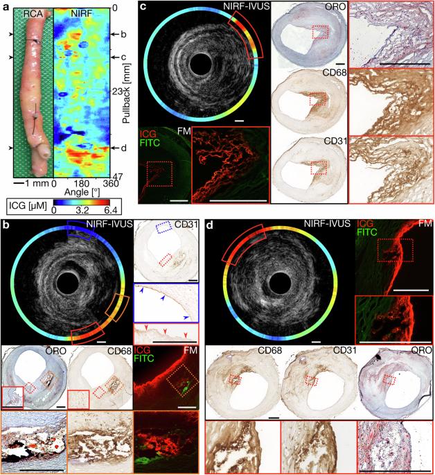
Indocyanine green (ICG)-enhanced intravascular near-infrared fluorescence (NIRF) imaging enhances the information obtained with intravascular ultrasound (IVUS) by visualizing pathobiological characteristics of atherosclerotic plaques. To advance our understanding of this hybrid method, we aimed to assess the potential of NIRF-IVUS to identify different stages of atheroma progression by characterizing ICG uptake in human pathological specimens. After excision, 15 human coronary specimens from 13 adult patients were ICG-perfused and imaged with NIRF-IVUS. All specimens were then histopathologically and immunohistochemically assessed. NIRF-IVUS imaging revealed colocalization of ICG-deposition to plaque areas of lipid accumulation, endothelial disruption, neovascularization and inflammation. Moreover, ICG concentrations were significantly higher in advanced coronary artery disease stages (p < 0.05) and correlated significantly to plaque macrophage burden (r = 0.67). Current intravascular methods fail to detect plaque biology. Thus, we demonstrate how human coronary atheroma stage can be assessed based on pathobiological characteristics uniquely captured by ICG-enhanced intravascular NIRF.

 求助内容:
求助内容: 应助结果提醒方式:


