{"title":"利用荧光寿命成像(FLIM)对复杂矿物基质中的微生物进行原位检测。","authors":"Yekaterina Chmykh, Jay L. Nadeau","doi":"10.1111/jmi.13264","DOIUrl":null,"url":null,"abstract":"<p>The utility of fluorescence lifetime imaging microscopy (FLIM) for identifying bacteria in complex mineral matrices was investigated. Baseline signals from unlabelled <i>Bacillus subtilis</i> and <i>Euglena gracilis</i>, and <i>Bacillus subtilis</i> labelled with SYTO 9 were obtained using two-photon excitation at 730, 750 and 800 nm, identifying characteristic lifetimes of photosynthetic pigments, unpigmented cellular autofluorescence, and SYTO 9. Labelled and unlabelled <i>B. subtilis</i> were seeded onto marble and gypsum samples containing endolithic photosynthetic cyanobacteria and the ability to distinguish cells from mineral autofluorescence and nonspecific dye staining was examined in parallel with ordinary multichannel confocal imaging. It was found that FLIM enabled discrimination of SYTO 9 labelled cells from background, but that the lifetime of SYTO 9 was shorter in cells on minerals than in pure culture under our conditions. Photosynthetic microorganisms were easily observed using both FLIM and confocal. Unlabelled, nonpigmented bacteria showed weak signals that were difficult to distinguish from background when minerals were present, though cellular autofluorescence consistent with NAD(P)H could be seen in pure cultures, and phasor analysis permitted detection on rocks. Gypsum and marble samples showed similar autofluorescence profiles, with little autofluorescence in the yellow-to-red range. Lifetime or time-gated imaging may prove a useful tool for environmental microbiology.</p><p><b>LAY DESCRIPTION</b>: The standard method of bacterial enumeration is to label the cells with a fluorescent dye and count them under high-power fluorescence microscopy. However, this can be difficult when the cells are embedded in soil and rock due to fluorescence from the surrounding minerals and dye binding to ambiguous features of the substrate. The use of fluorescence lifetime imaging (FLIM) can disambiguate these signals and allow for improved detection of bacteria in environmental samples.</p>","PeriodicalId":16484,"journal":{"name":"Journal of microscopy","volume":null,"pages":null},"PeriodicalIF":1.5000,"publicationDate":"2024-01-17","publicationTypes":"Journal Article","fieldsOfStudy":null,"isOpenAccess":false,"openAccessPdf":"https://onlinelibrary.wiley.com/doi/epdf/10.1111/jmi.13264","citationCount":"0","resultStr":"{\"title\":\"The use of fluorescence lifetime imaging (FLIM) for in situ microbial detection in complex mineral substrates\",\"authors\":\"Yekaterina Chmykh, Jay L. Nadeau\",\"doi\":\"10.1111/jmi.13264\",\"DOIUrl\":null,\"url\":null,\"abstract\":\"<p>The utility of fluorescence lifetime imaging microscopy (FLIM) for identifying bacteria in complex mineral matrices was investigated. Baseline signals from unlabelled <i>Bacillus subtilis</i> and <i>Euglena gracilis</i>, and <i>Bacillus subtilis</i> labelled with SYTO 9 were obtained using two-photon excitation at 730, 750 and 800 nm, identifying characteristic lifetimes of photosynthetic pigments, unpigmented cellular autofluorescence, and SYTO 9. Labelled and unlabelled <i>B. subtilis</i> were seeded onto marble and gypsum samples containing endolithic photosynthetic cyanobacteria and the ability to distinguish cells from mineral autofluorescence and nonspecific dye staining was examined in parallel with ordinary multichannel confocal imaging. It was found that FLIM enabled discrimination of SYTO 9 labelled cells from background, but that the lifetime of SYTO 9 was shorter in cells on minerals than in pure culture under our conditions. Photosynthetic microorganisms were easily observed using both FLIM and confocal. Unlabelled, nonpigmented bacteria showed weak signals that were difficult to distinguish from background when minerals were present, though cellular autofluorescence consistent with NAD(P)H could be seen in pure cultures, and phasor analysis permitted detection on rocks. Gypsum and marble samples showed similar autofluorescence profiles, with little autofluorescence in the yellow-to-red range. Lifetime or time-gated imaging may prove a useful tool for environmental microbiology.</p><p><b>LAY DESCRIPTION</b>: The standard method of bacterial enumeration is to label the cells with a fluorescent dye and count them under high-power fluorescence microscopy. However, this can be difficult when the cells are embedded in soil and rock due to fluorescence from the surrounding minerals and dye binding to ambiguous features of the substrate. The use of fluorescence lifetime imaging (FLIM) can disambiguate these signals and allow for improved detection of bacteria in environmental samples.</p>\",\"PeriodicalId\":16484,\"journal\":{\"name\":\"Journal of microscopy\",\"volume\":null,\"pages\":null},\"PeriodicalIF\":1.5000,\"publicationDate\":\"2024-01-17\",\"publicationTypes\":\"Journal Article\",\"fieldsOfStudy\":null,\"isOpenAccess\":false,\"openAccessPdf\":\"https://onlinelibrary.wiley.com/doi/epdf/10.1111/jmi.13264\",\"citationCount\":\"0\",\"resultStr\":null,\"platform\":\"Semanticscholar\",\"paperid\":null,\"PeriodicalName\":\"Journal of microscopy\",\"FirstCategoryId\":\"5\",\"ListUrlMain\":\"https://onlinelibrary.wiley.com/doi/10.1111/jmi.13264\",\"RegionNum\":4,\"RegionCategory\":\"工程技术\",\"ArticlePicture\":[],\"TitleCN\":null,\"AbstractTextCN\":null,\"PMCID\":null,\"EPubDate\":\"\",\"PubModel\":\"\",\"JCR\":\"Q3\",\"JCRName\":\"MICROSCOPY\",\"Score\":null,\"Total\":0}","platform":"Semanticscholar","paperid":null,"PeriodicalName":"Journal of microscopy","FirstCategoryId":"5","ListUrlMain":"https://onlinelibrary.wiley.com/doi/10.1111/jmi.13264","RegionNum":4,"RegionCategory":"工程技术","ArticlePicture":[],"TitleCN":null,"AbstractTextCN":null,"PMCID":null,"EPubDate":"","PubModel":"","JCR":"Q3","JCRName":"MICROSCOPY","Score":null,"Total":0}
The use of fluorescence lifetime imaging (FLIM) for in situ microbial detection in complex mineral substrates
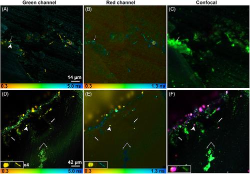
The utility of fluorescence lifetime imaging microscopy (FLIM) for identifying bacteria in complex mineral matrices was investigated. Baseline signals from unlabelled Bacillus subtilis and Euglena gracilis, and Bacillus subtilis labelled with SYTO 9 were obtained using two-photon excitation at 730, 750 and 800 nm, identifying characteristic lifetimes of photosynthetic pigments, unpigmented cellular autofluorescence, and SYTO 9. Labelled and unlabelled B. subtilis were seeded onto marble and gypsum samples containing endolithic photosynthetic cyanobacteria and the ability to distinguish cells from mineral autofluorescence and nonspecific dye staining was examined in parallel with ordinary multichannel confocal imaging. It was found that FLIM enabled discrimination of SYTO 9 labelled cells from background, but that the lifetime of SYTO 9 was shorter in cells on minerals than in pure culture under our conditions. Photosynthetic microorganisms were easily observed using both FLIM and confocal. Unlabelled, nonpigmented bacteria showed weak signals that were difficult to distinguish from background when minerals were present, though cellular autofluorescence consistent with NAD(P)H could be seen in pure cultures, and phasor analysis permitted detection on rocks. Gypsum and marble samples showed similar autofluorescence profiles, with little autofluorescence in the yellow-to-red range. Lifetime or time-gated imaging may prove a useful tool for environmental microbiology.
LAY DESCRIPTION: The standard method of bacterial enumeration is to label the cells with a fluorescent dye and count them under high-power fluorescence microscopy. However, this can be difficult when the cells are embedded in soil and rock due to fluorescence from the surrounding minerals and dye binding to ambiguous features of the substrate. The use of fluorescence lifetime imaging (FLIM) can disambiguate these signals and allow for improved detection of bacteria in environmental samples.
期刊介绍:
The Journal of Microscopy is the oldest journal dedicated to the science of microscopy and the only peer-reviewed publication of the Royal Microscopical Society. It publishes papers that report on the very latest developments in microscopy such as advances in microscopy techniques or novel areas of application. The Journal does not seek to publish routine applications of microscopy or specimen preparation even though the submission may otherwise have a high scientific merit.
The scope covers research in the physical and biological sciences and covers imaging methods using light, electrons, X-rays and other radiations as well as atomic force and near field techniques. Interdisciplinary research is welcome. Papers pertaining to microscopy are also welcomed on optical theory, spectroscopy, novel specimen preparation and manipulation methods and image recording, processing and analysis including dynamic analysis of living specimens.
Publication types include full papers, hot topic fast tracked communications and review articles. Authors considering submitting a review article should contact the editorial office first.

 求助内容:
求助内容: 应助结果提醒方式:


