David Pitcher, Magdalena W Sliwinska, Daniel Kaiser
{"title":"经颅磁刺激对外侧前额皮质的破坏增加了默认模式网络在命名面部表情时的神经活动。","authors":"David Pitcher, Magdalena W Sliwinska, Daniel Kaiser","doi":"10.1093/scan/nsad072","DOIUrl":null,"url":null,"abstract":"<p><p>Recognizing facial expressions is dependent on multiple brain networks specialized for different cognitive functions. In the current study, participants (N = 20) were scanned using functional magnetic resonance imaging (fMRI), while they performed a covert facial expression naming task. Immediately prior to scanning thetaburst transcranial magnetic stimulation (TMS) was delivered over the right lateral prefrontal cortex (PFC), or the vertex control site. A group whole-brain analysis revealed that TMS induced opposite effects in the neural responses across different brain networks. Stimulation of the right PFC (compared to stimulation of the vertex) decreased neural activity in the left lateral PFC but increased neural activity in three nodes of the default mode network (DMN): the right superior frontal gyrus, right angular gyrus and the bilateral middle cingulate gyrus. A region of interest analysis showed that TMS delivered over the right PFC reduced neural activity across all functionally localised face areas (including in the PFC) compared to TMS delivered over the vertex. These results suggest that visually recognizing facial expressions is dependent on the dynamic interaction of the face-processing network and the DMN. Our study also demonstrates the utility of combined TMS/fMRI studies for revealing the dynamic interactions between different functional brain networks.</p>","PeriodicalId":94208,"journal":{"name":"Social cognitive and affective neuroscience","volume":"18 1","pages":""},"PeriodicalIF":3.1000,"publicationDate":"2023-11-30","publicationTypes":"Journal Article","fieldsOfStudy":null,"isOpenAccess":false,"openAccessPdf":"https://www.ncbi.nlm.nih.gov/pmc/articles/PMC10695328/pdf/","citationCount":"0","resultStr":"{\"title\":\"TMS disruption of the lateral prefrontal cortex increases neural activity in the default mode network when naming facial expressions.\",\"authors\":\"David Pitcher, Magdalena W Sliwinska, Daniel Kaiser\",\"doi\":\"10.1093/scan/nsad072\",\"DOIUrl\":null,\"url\":null,\"abstract\":\"<p><p>Recognizing facial expressions is dependent on multiple brain networks specialized for different cognitive functions. In the current study, participants (N = 20) were scanned using functional magnetic resonance imaging (fMRI), while they performed a covert facial expression naming task. Immediately prior to scanning thetaburst transcranial magnetic stimulation (TMS) was delivered over the right lateral prefrontal cortex (PFC), or the vertex control site. A group whole-brain analysis revealed that TMS induced opposite effects in the neural responses across different brain networks. Stimulation of the right PFC (compared to stimulation of the vertex) decreased neural activity in the left lateral PFC but increased neural activity in three nodes of the default mode network (DMN): the right superior frontal gyrus, right angular gyrus and the bilateral middle cingulate gyrus. A region of interest analysis showed that TMS delivered over the right PFC reduced neural activity across all functionally localised face areas (including in the PFC) compared to TMS delivered over the vertex. These results suggest that visually recognizing facial expressions is dependent on the dynamic interaction of the face-processing network and the DMN. Our study also demonstrates the utility of combined TMS/fMRI studies for revealing the dynamic interactions between different functional brain networks.</p>\",\"PeriodicalId\":94208,\"journal\":{\"name\":\"Social cognitive and affective neuroscience\",\"volume\":\"18 1\",\"pages\":\"\"},\"PeriodicalIF\":3.1000,\"publicationDate\":\"2023-11-30\",\"publicationTypes\":\"Journal Article\",\"fieldsOfStudy\":null,\"isOpenAccess\":false,\"openAccessPdf\":\"https://www.ncbi.nlm.nih.gov/pmc/articles/PMC10695328/pdf/\",\"citationCount\":\"0\",\"resultStr\":null,\"platform\":\"Semanticscholar\",\"paperid\":null,\"PeriodicalName\":\"Social cognitive and affective neuroscience\",\"FirstCategoryId\":\"1085\",\"ListUrlMain\":\"https://doi.org/10.1093/scan/nsad072\",\"RegionNum\":0,\"RegionCategory\":null,\"ArticlePicture\":[],\"TitleCN\":null,\"AbstractTextCN\":null,\"PMCID\":null,\"EPubDate\":\"\",\"PubModel\":\"\",\"JCR\":\"\",\"JCRName\":\"\",\"Score\":null,\"Total\":0}","platform":"Semanticscholar","paperid":null,"PeriodicalName":"Social cognitive and affective neuroscience","FirstCategoryId":"1085","ListUrlMain":"https://doi.org/10.1093/scan/nsad072","RegionNum":0,"RegionCategory":null,"ArticlePicture":[],"TitleCN":null,"AbstractTextCN":null,"PMCID":null,"EPubDate":"","PubModel":"","JCR":"","JCRName":"","Score":null,"Total":0}
TMS disruption of the lateral prefrontal cortex increases neural activity in the default mode network when naming facial expressions.
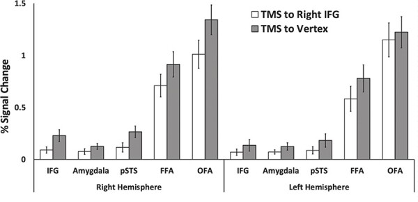
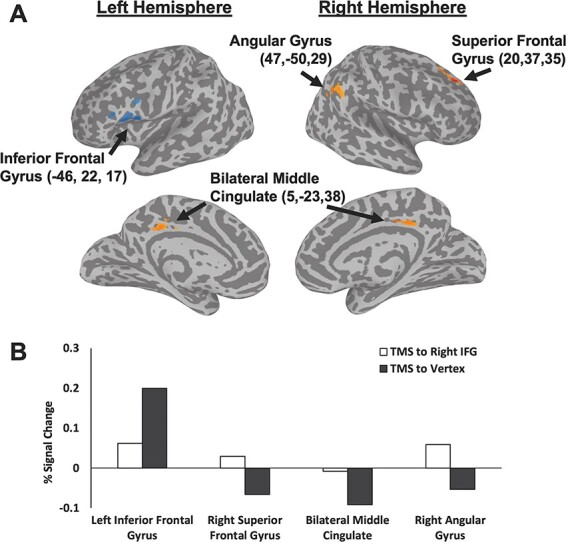
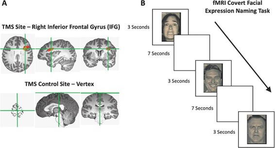
Recognizing facial expressions is dependent on multiple brain networks specialized for different cognitive functions. In the current study, participants (N = 20) were scanned using functional magnetic resonance imaging (fMRI), while they performed a covert facial expression naming task. Immediately prior to scanning thetaburst transcranial magnetic stimulation (TMS) was delivered over the right lateral prefrontal cortex (PFC), or the vertex control site. A group whole-brain analysis revealed that TMS induced opposite effects in the neural responses across different brain networks. Stimulation of the right PFC (compared to stimulation of the vertex) decreased neural activity in the left lateral PFC but increased neural activity in three nodes of the default mode network (DMN): the right superior frontal gyrus, right angular gyrus and the bilateral middle cingulate gyrus. A region of interest analysis showed that TMS delivered over the right PFC reduced neural activity across all functionally localised face areas (including in the PFC) compared to TMS delivered over the vertex. These results suggest that visually recognizing facial expressions is dependent on the dynamic interaction of the face-processing network and the DMN. Our study also demonstrates the utility of combined TMS/fMRI studies for revealing the dynamic interactions between different functional brain networks.

 求助内容:
求助内容: 应助结果提醒方式:


