Keiko Fukino, Kohsei Iida, Masahiro Tsutsumi, Joe Iwanaga, Keiichi Akita
{"title":"Evaluation of morphological features of palatopharyngeus insertion into the thyroid cartilage","authors":"Keiko Fukino, Kohsei Iida, Masahiro Tsutsumi, Joe Iwanaga, Keiichi Akita","doi":"10.1007/s12565-023-00709-7","DOIUrl":null,"url":null,"abstract":"<div><p>The attachment of the palatopharyngeus extended from the posterior end of the thyroid cartilage to the posterior margin of the inferior constrictor attachment that might contribute to successive swallowing movements. Laryngeal elevation is essential for proper swallowing and breathing. Recently, clinical research has demonstrated that the palatopharyngeus, a longitudinal muscle of the pharynx, is involved in the elevation of the larynx. However, the morphological relationship between the larynx and palatopharyngeus remains unclear. In the present study, we analyzed the attachment site and characteristics of the palatopharyngeus in the thyroid cartilage. We evaluated 14 halves of seven heads from Japanese cadavers (average age: 76.4 years); 12 halves, anatomically and two halves histologically. A part of the palatopharyngeus, which originated from the inferior aspect of the palatine aponeurosis, was attached to the inner and outer surfaces of the thyroid cartilage through collagen fibers. The attachment area extends from the posterior end of the thyroid cartilage to the posterior margin of the attachment site of the inferior constrictor. The palatopharyngeus may elevate the larynx with the suprahyoid muscles and contribute to successive movements of swallowing with surrounding muscles. Based on our findings and previous studies, palatopharyngeus with various muscle bundle directions may be essential for the coordination of continuous swallowing events.</p></div>","PeriodicalId":7816,"journal":{"name":"Anatomical Science International","volume":null,"pages":null},"PeriodicalIF":1.2000,"publicationDate":"2023-03-06","publicationTypes":"Journal Article","fieldsOfStudy":null,"isOpenAccess":false,"openAccessPdf":"","citationCount":"0","resultStr":null,"platform":"Semanticscholar","paperid":null,"PeriodicalName":"Anatomical Science International","FirstCategoryId":"3","ListUrlMain":"https://link.springer.com/article/10.1007/s12565-023-00709-7","RegionNum":4,"RegionCategory":"医学","ArticlePicture":[],"TitleCN":null,"AbstractTextCN":null,"PMCID":null,"EPubDate":"","PubModel":"","JCR":"Q3","JCRName":"ANATOMY & MORPHOLOGY","Score":null,"Total":0}
引用次数: 0
Abstract
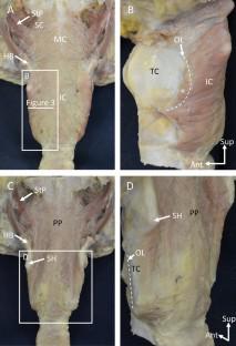
The attachment of the palatopharyngeus extended from the posterior end of the thyroid cartilage to the posterior margin of the inferior constrictor attachment that might contribute to successive swallowing movements. Laryngeal elevation is essential for proper swallowing and breathing. Recently, clinical research has demonstrated that the palatopharyngeus, a longitudinal muscle of the pharynx, is involved in the elevation of the larynx. However, the morphological relationship between the larynx and palatopharyngeus remains unclear. In the present study, we analyzed the attachment site and characteristics of the palatopharyngeus in the thyroid cartilage. We evaluated 14 halves of seven heads from Japanese cadavers (average age: 76.4 years); 12 halves, anatomically and two halves histologically. A part of the palatopharyngeus, which originated from the inferior aspect of the palatine aponeurosis, was attached to the inner and outer surfaces of the thyroid cartilage through collagen fibers. The attachment area extends from the posterior end of the thyroid cartilage to the posterior margin of the attachment site of the inferior constrictor. The palatopharyngeus may elevate the larynx with the suprahyoid muscles and contribute to successive movements of swallowing with surrounding muscles. Based on our findings and previous studies, palatopharyngeus with various muscle bundle directions may be essential for the coordination of continuous swallowing events.
期刊介绍:
The official English journal of the Japanese Association of Anatomists, Anatomical Science International (formerly titled Kaibogaku Zasshi) publishes original research articles dealing with morphological sciences.
Coverage in the journal includes molecular, cellular, histological and gross anatomical studies on humans and on normal and experimental animals, as well as functional morphological, biochemical, physiological and behavioral studies if they include morphological analysis.

 求助内容:
求助内容: 应助结果提醒方式:


