Comparison of Retinal Microvascular Vascular Density Between Adolescents With and Without Simple Myopia Using Optical Coherence Tomography Angiography.
{"title":"Comparison of Retinal Microvascular Vascular Density Between Adolescents With and Without Simple Myopia Using Optical Coherence Tomography Angiography.","authors":"Kemal Bayrakçeken","doi":"10.5152/eurasianjmed.2023.22278","DOIUrl":null,"url":null,"abstract":"<p><strong>Objective: </strong>The aim of this study is to investigate whether there is a difference in retinal microvascularization between adolescents with and without simple myopia using optical coherence tomography angiography.</p><p><strong>Materials and methods: </strong>Thirty-four eyes of 34 patients aged 12-18 years diagnosed with school-age simple myopia (0-6 diopters), and 34 eyes of 34 healthy controls of similar ages were included in this retrospective study. The ocular, optical coherence tomography, and optical coherence tomography angiography findings of the participants were recorded.</p><p><strong>Results: </strong>The simple myopia group had statistically thicker inferior ganglion cell complex thicknesses compared to the controls (P =.038). The macular map values did not statistically significantly differ between the 2 groups. The foveal avascular zone area (P =.038) and circularity index (P =.022) values were statistically lower in the simple myopia group compared to the control group. The superficial capillary plexus outer and inner ring vessel density (%) (superior and nasal) showed statistically significant differences (outer ring superior/ nasal P=.004/P = .037; inner ring superior/nasal P =.014/P= .046).</p><p><strong>Conclusion: </strong>Similar to high myopia, vascular density in the macula decreases as the axial length and spherical equivalent increase in simple myopia.</p>","PeriodicalId":53592,"journal":{"name":"Eurasian Journal of Medicine","volume":null,"pages":null},"PeriodicalIF":0.9000,"publicationDate":"2023-02-01","publicationTypes":"Journal Article","fieldsOfStudy":null,"isOpenAccess":false,"openAccessPdf":"https://www.ncbi.nlm.nih.gov/pmc/articles/PMC10081140/pdf/","citationCount":"1","resultStr":null,"platform":"Semanticscholar","paperid":null,"PeriodicalName":"Eurasian Journal of Medicine","FirstCategoryId":"1085","ListUrlMain":"https://doi.org/10.5152/eurasianjmed.2023.22278","RegionNum":0,"RegionCategory":null,"ArticlePicture":[],"TitleCN":null,"AbstractTextCN":null,"PMCID":null,"EPubDate":"","PubModel":"","JCR":"Q3","JCRName":"MEDICINE, GENERAL & INTERNAL","Score":null,"Total":0}
引用次数: 1
Abstract
Objective: The aim of this study is to investigate whether there is a difference in retinal microvascularization between adolescents with and without simple myopia using optical coherence tomography angiography.
Materials and methods: Thirty-four eyes of 34 patients aged 12-18 years diagnosed with school-age simple myopia (0-6 diopters), and 34 eyes of 34 healthy controls of similar ages were included in this retrospective study. The ocular, optical coherence tomography, and optical coherence tomography angiography findings of the participants were recorded.
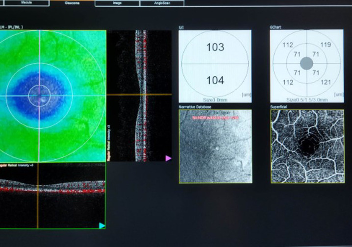
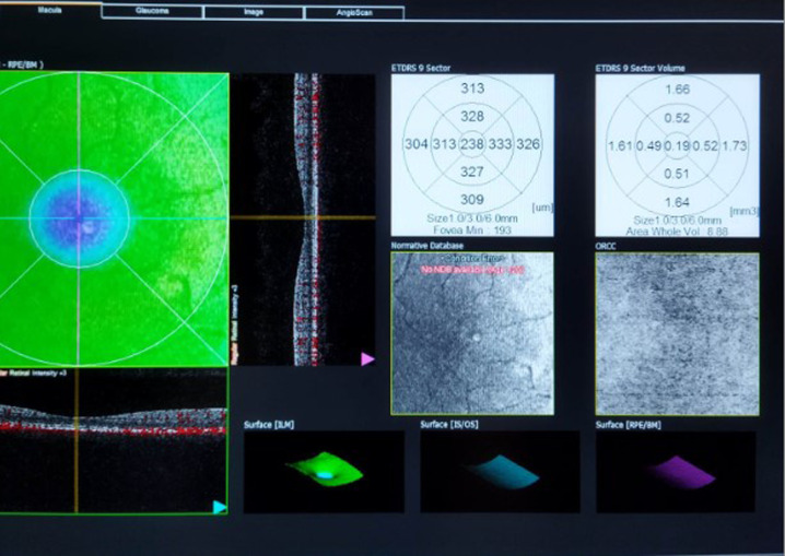
Results: The simple myopia group had statistically thicker inferior ganglion cell complex thicknesses compared to the controls (P =.038). The macular map values did not statistically significantly differ between the 2 groups. The foveal avascular zone area (P =.038) and circularity index (P =.022) values were statistically lower in the simple myopia group compared to the control group. The superficial capillary plexus outer and inner ring vessel density (%) (superior and nasal) showed statistically significant differences (outer ring superior/ nasal P=.004/P = .037; inner ring superior/nasal P =.014/P= .046).
Conclusion: Similar to high myopia, vascular density in the macula decreases as the axial length and spherical equivalent increase in simple myopia.
期刊介绍:
Eurasian Journal of Medicine (Eurasian J Med) is an international, scientific, open access periodical published by independent, unbiased, and triple-blinded peer-review principles. The journal is the official publication of Atatürk University School of Medicine and published triannually in February, June, and October. The publication language of the journal is English. The aim of the Eurasian Journal of Medicine is to publish original research papers of the highest scientific and clinical value in all medical fields. The Eurasian J Med also includes reviews, editorial short notes and letters to the editor that either as a comment related to recently published articles in our journal or as a case report. The target audience of the journal includes researchers, physicians and healthcare professionals who are interested or working in in all medical disciplines.


 求助内容:
求助内容: 应助结果提醒方式:


