Management of a Bilateral Post-Uveitic Complex Glaucoma with Pupillary Block, Rupture of the Anterior Lens Capsule, and Malignant Glaucoma following Laser Peripheral Iridotomies: Case Report and Literature Review.
Khaled El Matri, Dhouha Gouider, Rim Limaiem, Ahmed Chebil, Meher Henchiri, Yousra Falfoul, Leila El Matri
{"title":"Management of a Bilateral Post-Uveitic Complex Glaucoma with Pupillary Block, Rupture of the Anterior Lens Capsule, and Malignant Glaucoma following Laser Peripheral Iridotomies: Case Report and Literature Review.","authors":"Khaled El Matri, Dhouha Gouider, Rim Limaiem, Ahmed Chebil, Meher Henchiri, Yousra Falfoul, Leila El Matri","doi":"10.4103/joco.joco_3_22","DOIUrl":null,"url":null,"abstract":"<p><strong>Purpose: </strong>To report a case of a bilateral complex uveitic glaucoma (UG) with pupillary block, rupture of the anterior lens capsule, and malignant glaucoma in a young high-myopic patient and to report anterior segment optical coherence tomography (AS-OCT) findings initially and following surgery.</p><p><strong>Methods: </strong>A 21-year-old high-myopic woman who had a history of anterior uveitis with extensive posterior synechiae, presented with acute bilateral ocular pain, redness, and blurred vision following bilateral Nd: YAG laser peripheral iridotomy (LPI).</p><p><strong>Results: </strong>Visual acuity was limited to light perception in both eyes (OU), with a flat anterior chamber (AC) and anterior luxation of lens fragments. Intraocular pressure (IOP) was over 60 mmHg OU. AS-OCT showed closed angles and hyperreflective heterogeneous material within the flat AC. The iris and lens fragments were plated against the corneal endothelium OU. We performed an urgent pars plana vitrectomy associated with lensectomy. It was uneventful in OU. Repeated AS-OCT revealed a deep AC, widely open angles, and aphakia. IOP was lowered to 9 mmHg and visual acuity improved to 5/10 in OU.</p><p><strong>Conclusion: </strong>Performing LPI might be harmful in the presence of UG with extensive posterior synechia, resulting in complex mechanism glaucoma with aqueous misdirection syndrome associated with a pupillary block due to anterior lens luxation, even in high-myopic eyes. Nd: YAG LPI should not be performed simultaneously in OU, especially in pathologic eyes, to prevent bilateral vision-threatening complications. AS-OCT was of great help, allowing easy and detailed ultrastructural assessment of the ACs, and iridocorneal angles before and after surgery.</p>","PeriodicalId":15423,"journal":{"name":"Journal of Current Ophthalmology","volume":null,"pages":null},"PeriodicalIF":1.2000,"publicationDate":"2022-07-26","publicationTypes":"Journal Article","fieldsOfStudy":null,"isOpenAccess":false,"openAccessPdf":"https://ftp.ncbi.nlm.nih.gov/pub/pmc/oa_pdf/0f/84/JCO-34-260.PMC9487004.pdf","citationCount":"0","resultStr":null,"platform":"Semanticscholar","paperid":null,"PeriodicalName":"Journal of Current Ophthalmology","FirstCategoryId":"1085","ListUrlMain":"https://doi.org/10.4103/joco.joco_3_22","RegionNum":0,"RegionCategory":null,"ArticlePicture":[],"TitleCN":null,"AbstractTextCN":null,"PMCID":null,"EPubDate":"2022/4/1 0:00:00","PubModel":"eCollection","JCR":"Q3","JCRName":"OPHTHALMOLOGY","Score":null,"Total":0}
引用次数: 0
Abstract
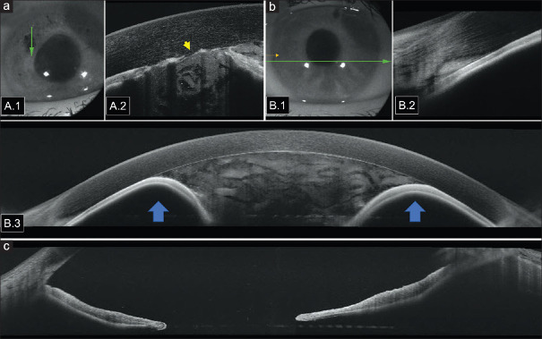
Purpose: To report a case of a bilateral complex uveitic glaucoma (UG) with pupillary block, rupture of the anterior lens capsule, and malignant glaucoma in a young high-myopic patient and to report anterior segment optical coherence tomography (AS-OCT) findings initially and following surgery.
Methods: A 21-year-old high-myopic woman who had a history of anterior uveitis with extensive posterior synechiae, presented with acute bilateral ocular pain, redness, and blurred vision following bilateral Nd: YAG laser peripheral iridotomy (LPI).
Results: Visual acuity was limited to light perception in both eyes (OU), with a flat anterior chamber (AC) and anterior luxation of lens fragments. Intraocular pressure (IOP) was over 60 mmHg OU. AS-OCT showed closed angles and hyperreflective heterogeneous material within the flat AC. The iris and lens fragments were plated against the corneal endothelium OU. We performed an urgent pars plana vitrectomy associated with lensectomy. It was uneventful in OU. Repeated AS-OCT revealed a deep AC, widely open angles, and aphakia. IOP was lowered to 9 mmHg and visual acuity improved to 5/10 in OU.
Conclusion: Performing LPI might be harmful in the presence of UG with extensive posterior synechia, resulting in complex mechanism glaucoma with aqueous misdirection syndrome associated with a pupillary block due to anterior lens luxation, even in high-myopic eyes. Nd: YAG LPI should not be performed simultaneously in OU, especially in pathologic eyes, to prevent bilateral vision-threatening complications. AS-OCT was of great help, allowing easy and detailed ultrastructural assessment of the ACs, and iridocorneal angles before and after surgery.
期刊介绍:
Peer Review under the responsibility of Iranian Society of Ophthalmology Journal of Current Ophthalmology, the official publication of the Iranian Society of Ophthalmology, is a peer-reviewed, open-access, scientific journal that welcomes high quality original articles related to vision science and all fields of ophthalmology. Journal of Current Ophthalmology is the continuum of Iranian Journal of Ophthalmology published since 1969.

 求助内容:
求助内容: 应助结果提醒方式:


