Implants with a Laser-microgrooved Collar Placed in Grafted Posterior Maxillary Extraction Sockets and in Crestally Grafted Sinuses: a 5-Year Multicentre Retrospective Study.
{"title":"Implants with a Laser-microgrooved Collar Placed in Grafted Posterior Maxillary Extraction Sockets and in Crestally Grafted Sinuses: a 5-Year Multicentre Retrospective Study.","authors":"Renzo Guarnieri, Luca Savio, Alessandro Bermonds, Luca Testarelli","doi":"10.5037/jomr.2020.11402","DOIUrl":null,"url":null,"abstract":"<p><strong>Objectives: </strong>The aim of this retrospective multicentre cohort study was to compare clinical outcomes, soft tissues conditions and differences in marginal bone loss between implants with a laser-microgrooved collar placed in posterior maxillary extraction sockets grafted by 4 to 5 months, and in posterior maxillary pristine bone (spontaneously healed posterior maxillary extraction sockets) by means of osteotome-mediated sinus floor elevation, over a period of 5 years after functional loading.</p><p><strong>Material and methods: </strong>Patients of Group 1 underwent extractions with sockets preservation using porcine-derived bone, covered with collagen membrane. Group 2 underwent extractions without socket preservation. Patients of Group 1 received implants in grafted sites, and Group 2 received implants in spontaneously healed bone using a maxillary sinus lift with crestal approach.</p><p><strong>Results: </strong>Over the observation period, the overall clinical success rate in Group 1 and Group 2 was 98% and 100%, respectively, with no differences between the procedures and implants used. Cumulative radiographic marginal bone loss ranged from 0.03 to 0.39 mm after 60 months of functional loading. There were no statistically significant differences in marginal bone loss between short and standard-length implants placed in grafted extraction sockets and in pristine bone.</p><p><strong>Conclusions: </strong>Short and standard implants with a laser-microgrooved collar, placed in posterior maxillary extraction sockets grafted by 4 to 5 months, and in posterior maxillary pristine bone (spontaneously healed posterior maxillary extraction sockets) by means of osteotome-mediated sinus floor elevation, exhibited no statistical difference in success rate, clinical parameters and marginal bone loss.</p>","PeriodicalId":230885,"journal":{"name":"Journal of Oral & Maxillofacial Research","volume":" ","pages":"e2"},"PeriodicalIF":0.0000,"publicationDate":"2020-12-31","publicationTypes":"Journal Article","fieldsOfStudy":null,"isOpenAccess":false,"openAccessPdf":"https://ftp.ncbi.nlm.nih.gov/pub/pmc/oa_pdf/36/ec/jomr-11-e2.PMC7875103.pdf","citationCount":"1","resultStr":null,"platform":"Semanticscholar","paperid":null,"PeriodicalName":"Journal of Oral & Maxillofacial Research","FirstCategoryId":"1085","ListUrlMain":"https://doi.org/10.5037/jomr.2020.11402","RegionNum":0,"RegionCategory":null,"ArticlePicture":[],"TitleCN":null,"AbstractTextCN":null,"PMCID":null,"EPubDate":"2020/10/1 0:00:00","PubModel":"eCollection","JCR":"","JCRName":"","Score":null,"Total":0}
引用次数: 1
Abstract
Objectives: The aim of this retrospective multicentre cohort study was to compare clinical outcomes, soft tissues conditions and differences in marginal bone loss between implants with a laser-microgrooved collar placed in posterior maxillary extraction sockets grafted by 4 to 5 months, and in posterior maxillary pristine bone (spontaneously healed posterior maxillary extraction sockets) by means of osteotome-mediated sinus floor elevation, over a period of 5 years after functional loading.
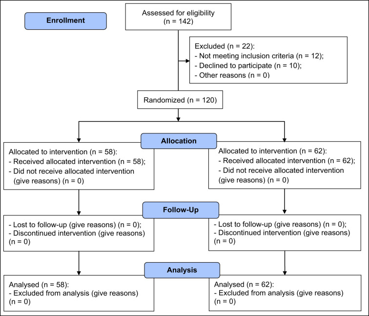
Material and methods: Patients of Group 1 underwent extractions with sockets preservation using porcine-derived bone, covered with collagen membrane. Group 2 underwent extractions without socket preservation. Patients of Group 1 received implants in grafted sites, and Group 2 received implants in spontaneously healed bone using a maxillary sinus lift with crestal approach.
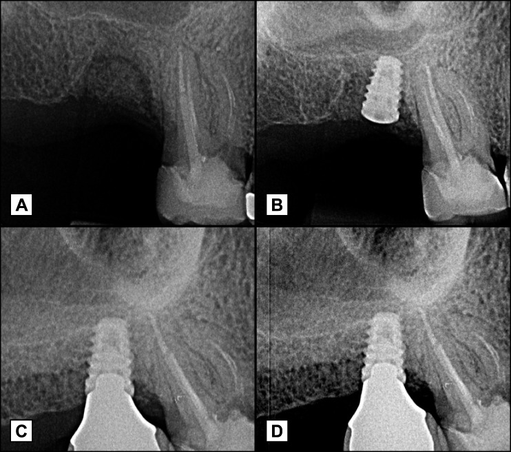
Results: Over the observation period, the overall clinical success rate in Group 1 and Group 2 was 98% and 100%, respectively, with no differences between the procedures and implants used. Cumulative radiographic marginal bone loss ranged from 0.03 to 0.39 mm after 60 months of functional loading. There were no statistically significant differences in marginal bone loss between short and standard-length implants placed in grafted extraction sockets and in pristine bone.
Conclusions: Short and standard implants with a laser-microgrooved collar, placed in posterior maxillary extraction sockets grafted by 4 to 5 months, and in posterior maxillary pristine bone (spontaneously healed posterior maxillary extraction sockets) by means of osteotome-mediated sinus floor elevation, exhibited no statistical difference in success rate, clinical parameters and marginal bone loss.


 求助内容:
求助内容: 应助结果提醒方式:


