Detailed Analysis of Fetal Malformations of the Supratentorial Structures of the Brain in High-Risk Pregnancies at 12-14 Gestational Weeks by Transvaginal 3D Ultrasound Examination.
IF 1.6 Q3 RADIOLOGY, NUCLEAR MEDICINE & MEDICAL IMAGING
Reinhard Altmann, Iris Scharnreitner, Sabine Enengl, Patrick Stelzl, Peter Oppelt, Elisabeth Reiter
{"title":"Detailed Analysis of Fetal Malformations of the Supratentorial Structures of the Brain in High-Risk Pregnancies at 12-14 Gestational Weeks by Transvaginal 3D Ultrasound Examination.","authors":"Reinhard Altmann, Iris Scharnreitner, Sabine Enengl, Patrick Stelzl, Peter Oppelt, Elisabeth Reiter","doi":"10.1055/a-2422-8443","DOIUrl":null,"url":null,"abstract":"<p><p><b>Purpose</b> To detect sonographic abnormalities of the supratentorial structures of the brain - future cavum septum pellucidum, cavum velum interpositum, third ventricle, ganglionic eminence and thalamus/hypothalamus - in fetuses with a crown-rump length of 45-84 mm in high-risk pregnancies. <b>Materials and Methods</b> This study presents the retrospective analysis of transvaginally recorded 3D volumes of the fetal brain of 64 fetuses whose mothers consulted our ambulatory department for fetomaternal medicine for organic and/or genetic changes of their fetuses at GW 12-14. For this study we selected fetuses with 3D volume blocks of the fetal brain at best sonographic quality enabling detailed analysis and measurement of the supratentorial brain structures to correlate the results with the results of genetic analysis, ultrasound controls in later weeks of pregnancy, and fetal outcome. <b>Results</b> Of 44 fetuses with genetic changes and 20 fetuses with syndromic changes, structural brain changes were found in 27 fetuses, analyzed by correlating the brain structures with the recently published structures of the brain at gestational week 12-14 in early pregnancy, presenting new details of early pathological brain development - migration disorders, milder variants of holoprosencephaly (lobar, MIH), corpus callosum agenesis, for the first time in early pregnancy. <b>Conclusion</b> Supratentorial defects of the brain can be detected and analyzed in GW 12-14 in detail by direct analysis of sonopathology and visualization of pathological measurements using transvaginal 3D sonography in high quality.</p>","PeriodicalId":44852,"journal":{"name":"Ultrasound International Open","volume":"10 ","pages":"a24228443"},"PeriodicalIF":1.6000,"publicationDate":"2024-12-16","publicationTypes":"Journal Article","fieldsOfStudy":null,"isOpenAccess":false,"openAccessPdf":"https://www.ncbi.nlm.nih.gov/pmc/articles/PMC11863995/pdf/","citationCount":"0","resultStr":null,"platform":"Semanticscholar","paperid":null,"PeriodicalName":"Ultrasound International Open","FirstCategoryId":"1085","ListUrlMain":"https://doi.org/10.1055/a-2422-8443","RegionNum":0,"RegionCategory":null,"ArticlePicture":[],"TitleCN":null,"AbstractTextCN":null,"PMCID":null,"EPubDate":"2024/1/1 0:00:00","PubModel":"eCollection","JCR":"Q3","JCRName":"RADIOLOGY, NUCLEAR MEDICINE & MEDICAL IMAGING","Score":null,"Total":0}
引用次数: 0
Abstract
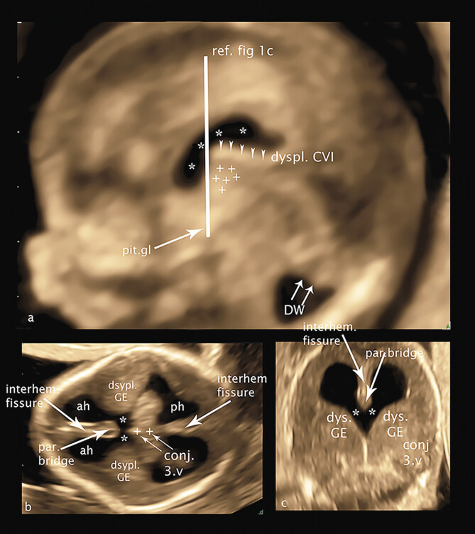
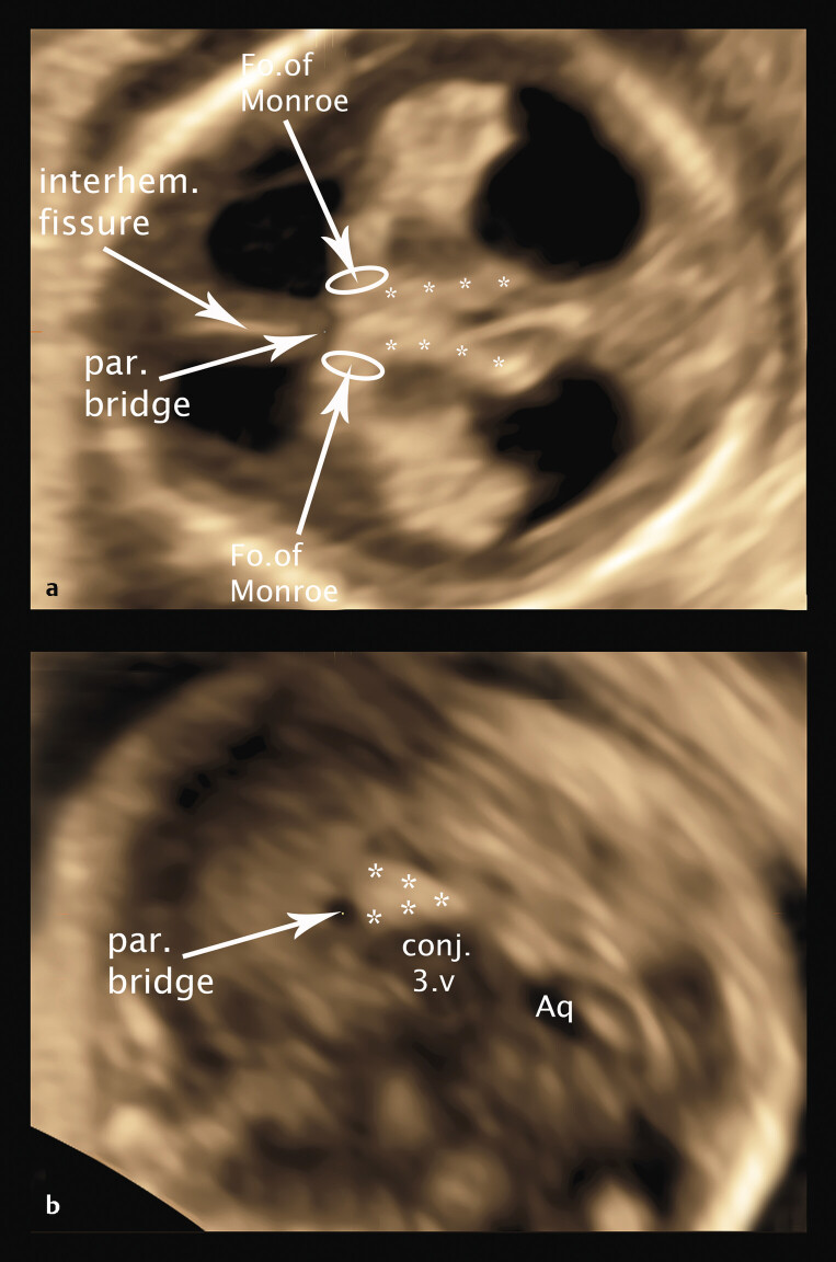
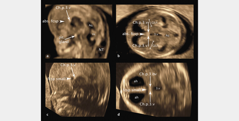
Purpose To detect sonographic abnormalities of the supratentorial structures of the brain - future cavum septum pellucidum, cavum velum interpositum, third ventricle, ganglionic eminence and thalamus/hypothalamus - in fetuses with a crown-rump length of 45-84 mm in high-risk pregnancies. Materials and Methods This study presents the retrospective analysis of transvaginally recorded 3D volumes of the fetal brain of 64 fetuses whose mothers consulted our ambulatory department for fetomaternal medicine for organic and/or genetic changes of their fetuses at GW 12-14. For this study we selected fetuses with 3D volume blocks of the fetal brain at best sonographic quality enabling detailed analysis and measurement of the supratentorial brain structures to correlate the results with the results of genetic analysis, ultrasound controls in later weeks of pregnancy, and fetal outcome. Results Of 44 fetuses with genetic changes and 20 fetuses with syndromic changes, structural brain changes were found in 27 fetuses, analyzed by correlating the brain structures with the recently published structures of the brain at gestational week 12-14 in early pregnancy, presenting new details of early pathological brain development - migration disorders, milder variants of holoprosencephaly (lobar, MIH), corpus callosum agenesis, for the first time in early pregnancy. Conclusion Supratentorial defects of the brain can be detected and analyzed in GW 12-14 in detail by direct analysis of sonopathology and visualization of pathological measurements using transvaginal 3D sonography in high quality.

 求助内容:
求助内容: 应助结果提醒方式:


