Non-invasive imaging biomarkers in chronic liver disease
IF 3.2
3区 医学
Q1 RADIOLOGY, NUCLEAR MEDICINE & MEDICAL IMAGING
引用次数: 0
Abstract
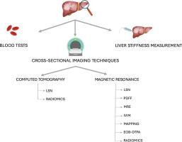
Chronic liver disease (CLD) is a global and worldwide clinical challenge, considering that different underlying liver entities can lead to hepatic dysfunction. In the past, blood tests and clinical evaluation were the main noninvasive tools used to detect, diagnose and follow-up patients with CLD; in case of clinical suspicion of CLD or unclear diagnosis, liver biopsy has been considered as the reference standard to rule out different chronic liver conditions. Nowadays, noninvasive tests have gained a central role in the clinical pathway. Particularly, liver stiffness measurement (LSM) and cross-sectional imaging techniques can provide transversal information to clinicians, helping them to correctly manage, treat and follow patients during time. Cross-sectional imaging techniques, namely computed tomography (CT) and magnetic resonance imaging (MRI), have plenty of potential. Both techniques allow to compute the liver surface nodularity (LSN), associated with CLDs and risk of decompensation. MRI can also help quantify fatty liver infiltration, mainly with the proton density fat fraction (PDFF) sequences, and detect and quantify fibrosis, especially thanks to elastography (MRE). Advanced techniques, such as intravoxel incoherent motion (IVIM), T1- and T2- mapping are promising tools for detecting fibrosis deposition. Furthermore, the injection of hepatobiliary contrast agents has gained an important role not only in liver lesion characterization but also in assessing liver function, especially in CLDs. Finally, the broad development of radiomics signatures, applied to CT and MR, can be considered the next future approach to CLDs. The aim of this review is to provide a comprehensive overview of the current advancements and applications of both invasive and noninvasive imaging techniques in the evaluation and management of CLD.

慢性肝病的无创成像生物标志物
慢性肝病(CLD)是一项全球性、世界性的临床挑战,因为不同的潜在肝脏实体都可能导致肝功能异常。过去,血液化验和临床评估是用于检测、诊断和随访慢性肝病患者的主要无创工具;在临床怀疑慢性肝病或诊断不明确的情况下,肝活检一直被视为排除不同慢性肝病的参考标准。如今,无创检测已在临床路径中占据重要地位。尤其是肝脏硬度测量(LSM)和横断面成像技术可以为临床医生提供横向信息,帮助他们正确管理、治疗和随访患者。横断面成像技术,即计算机断层扫描(CT)和磁共振成像(MRI),具有很大的潜力。这两种技术都可以计算肝表面结节度(LSN),这与慢性肝病和肝功能失代偿风险有关。核磁共振成像还能帮助量化脂肪肝浸润,主要是通过质子密度脂肪分数(PDFF)序列,以及检测和量化肝纤维化,特别是通过弹性成像(MRE)。先进的技术,如体内非相干运动(IVIM)、T1-和T2-映射,是检测纤维化沉积的有前途的工具。此外,注射肝胆造影剂不仅在肝脏病变定性方面,而且在评估肝功能方面,尤其是在慢性肝病方面,都发挥了重要作用。最后,应用于 CT 和 MR 的放射组学特征的广泛发展可被视为治疗 CLD 的下一个未来方法。本综述旨在全面概述目前有创和无创成像技术在评估和管理慢性肝病方面的进展和应用。
本文章由计算机程序翻译,如有差异,请以英文原文为准。
求助全文
约1分钟内获得全文
求助全文
来源期刊
CiteScore
6.70
自引率
3.00%
发文量
398
审稿时长
42 days
期刊介绍:
European Journal of Radiology is an international journal which aims to communicate to its readers, state-of-the-art information on imaging developments in the form of high quality original research articles and timely reviews on current developments in the field.
Its audience includes clinicians at all levels of training including radiology trainees, newly qualified imaging specialists and the experienced radiologist. Its aim is to inform efficient, appropriate and evidence-based imaging practice to the benefit of patients worldwide.

 求助内容:
求助内容: 应助结果提醒方式:


