Fatiguing exercise reduces cellular passive Young's modulus in human vastus lateralis muscle

IF 2.6 4区 医学 Q2 PHYSIOLOGY
Grace E. Privett, Austin W. Ricci, Larry L. David, Karen Wiedenfeld Needham, Yong How Tan, Karina H. Nakayama, Damien M. Callahan
{"title":"Fatiguing exercise reduces cellular passive Young's modulus in human vastus lateralis muscle","authors":"Grace E. Privett,&nbsp;Austin W. Ricci,&nbsp;Larry L. David,&nbsp;Karen Wiedenfeld Needham,&nbsp;Yong How Tan,&nbsp;Karina H. Nakayama,&nbsp;Damien M. Callahan","doi":"10.1113/EP092072","DOIUrl":null,"url":null,"abstract":"<div>\n \n <section>\n \n \n <p>Previous studies demonstrated that acute fatiguing exercise transiently reduces whole-muscle stiffness, which might contribute to increased risk of injury and impaired contractile performance. We sought to elucidate potential intracellular mechanisms underlying these reductions. To that end, the cellular passive Young's modulus was measured in muscle fibres from healthy, young males and females. Eight volunteers (four male and four female) completed unilateral, repeated maximal voluntary knee extensions until task failure, immediately followed by bilateral percutaneous needle muscle biopsy of the post-fatigued followed by the non-fatigued control vastus lateralis. Muscle samples were processed for mechanical assessment and separately for imaging and phosphoproteomics. Fibres were passively (pCa 8.0) stretched incrementally to 156% of initial sarcomere length to assess Young's modulus, calculated as the slope of the resulting stress–strain curve at short (sarcomere length = 2.4–3.0 µm) and long (sarcomere length = 3.2–3.8 µm) lengths. Titin phosphorylation was assessed by liquid chromatography followed by high-resolution mass spectrometry. The passive modulus was significantly reduced in post-fatigued versus control fibres from male, but not female, participants. Post-fatigued samples showed altered phosphorylation of five serine residues (four located within the elastic region of titin) but did not exhibit altered active tension or sarcomere ultrastructure. Collectively, these results suggest that acute fatigue is sufficient to alter phosphorylation of skeletal titin in multiple locations. We also found reductions in the passive modulus, consistent with prior reports in the literature investigating striated muscle stiffness. These results provide mechanistic insight contributing to the understanding of dynamic regulation of whole-muscle tissue mechanics in vivo.</p>\n </section>\n \n <section>\n \n <h3> Highlights</h3>\n \n <div>\n <ul>\n \n <li>\n <p><b>What is the central question of this study?</b></p>\n \n <p>Previous studies have shown that skeletal muscle stiffness is reduced following a single bout of fatiguing exercise in whole muscle, but it is not known whether these changes manifest at the cellular level, and their potential mechanisms remain unexplored.</p>\n </li>\n \n <li>\n <p><b>What is the main finding and its importance?</b></p>\n \n <p>Fatiguing exercise reduces cellular stiffness in skeletal muscle from males but not females, suggesting that fatigue alters tissue compliance in a sex-dependent manner. The phosphorylation status of titin, a potential mediator of skeletal muscle cellular stiffness, is modified by fatiguing exercise.</p>\n </li>\n \n <li>Previous studies have shown that passive skeletal muscle stiffness is reduced following a single bout of fatiguing exercise.</li>\n \n <li>Lower muscle passive stiffness following fatiguing exercise might increase risk for soft-tissue injury; however, the underlying mechanisms of this change are unclear.</li>\n \n <li>Our findings show that fatiguing exercise reduces the passive Young's modulus in skeletal muscle cells from males but not females, suggesting that intracellular proteins contribute to reduced muscle stiffness following repeated loading to task failure in a sex-dependent manner.</li>\n \n <li>The phosphorylation status of the intracellular protein titin is modified by fatiguing exercise in a way that might contribute to altered muscle stiffness after fatiguing exercise.</li>\n \n <li>These results provide important mechanistic insight that might help to explain why biological sex impacts the risk for soft-tissue injury with repeated or high-intensity mechanical loading in athletes and the risk of falls in older adults.</li>\n </ul>\n </div>\n </section>\n </div>","PeriodicalId":12092,"journal":{"name":"Experimental Physiology","volume":"109 11","pages":"1922-1937"},"PeriodicalIF":2.6000,"publicationDate":"2024-08-20","publicationTypes":"Journal Article","fieldsOfStudy":null,"isOpenAccess":false,"openAccessPdf":"https://onlinelibrary.wiley.com/doi/epdf/10.1113/EP092072","citationCount":"0","resultStr":null,"platform":"Semanticscholar","paperid":null,"PeriodicalName":"Experimental Physiology","FirstCategoryId":"3","ListUrlMain":"https://onlinelibrary.wiley.com/doi/10.1113/EP092072","RegionNum":4,"RegionCategory":"医学","ArticlePicture":[],"TitleCN":null,"AbstractTextCN":null,"PMCID":null,"EPubDate":"","PubModel":"","JCR":"Q2","JCRName":"PHYSIOLOGY","Score":null,"Total":0}
引用次数: 0

Abstract

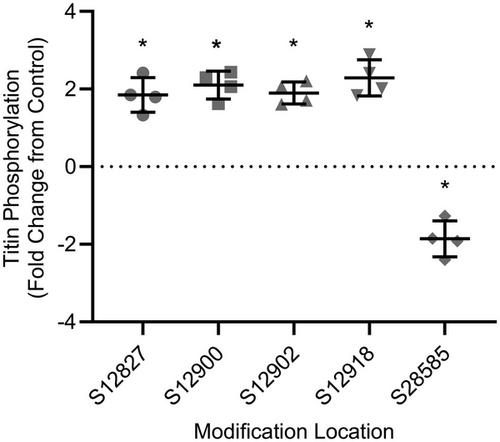
Previous studies demonstrated that acute fatiguing exercise transiently reduces whole-muscle stiffness, which might contribute to increased risk of injury and impaired contractile performance. We sought to elucidate potential intracellular mechanisms underlying these reductions. To that end, the cellular passive Young's modulus was measured in muscle fibres from healthy, young males and females. Eight volunteers (four male and four female) completed unilateral, repeated maximal voluntary knee extensions until task failure, immediately followed by bilateral percutaneous needle muscle biopsy of the post-fatigued followed by the non-fatigued control vastus lateralis. Muscle samples were processed for mechanical assessment and separately for imaging and phosphoproteomics. Fibres were passively (pCa 8.0) stretched incrementally to 156% of initial sarcomere length to assess Young's modulus, calculated as the slope of the resulting stress–strain curve at short (sarcomere length = 2.4–3.0 µm) and long (sarcomere length = 3.2–3.8 µm) lengths. Titin phosphorylation was assessed by liquid chromatography followed by high-resolution mass spectrometry. The passive modulus was significantly reduced in post-fatigued versus control fibres from male, but not female, participants. Post-fatigued samples showed altered phosphorylation of five serine residues (four located within the elastic region of titin) but did not exhibit altered active tension or sarcomere ultrastructure. Collectively, these results suggest that acute fatigue is sufficient to alter phosphorylation of skeletal titin in multiple locations. We also found reductions in the passive modulus, consistent with prior reports in the literature investigating striated muscle stiffness. These results provide mechanistic insight contributing to the understanding of dynamic regulation of whole-muscle tissue mechanics in vivo.

Highlights

  • What is the central question of this study?

    Previous studies have shown that skeletal muscle stiffness is reduced following a single bout of fatiguing exercise in whole muscle, but it is not known whether these changes manifest at the cellular level, and their potential mechanisms remain unexplored.

  • What is the main finding and its importance?

    Fatiguing exercise reduces cellular stiffness in skeletal muscle from males but not females, suggesting that fatigue alters tissue compliance in a sex-dependent manner. The phosphorylation status of titin, a potential mediator of skeletal muscle cellular stiffness, is modified by fatiguing exercise.

  • Previous studies have shown that passive skeletal muscle stiffness is reduced following a single bout of fatiguing exercise.
  • Lower muscle passive stiffness following fatiguing exercise might increase risk for soft-tissue injury; however, the underlying mechanisms of this change are unclear.
  • Our findings show that fatiguing exercise reduces the passive Young's modulus in skeletal muscle cells from males but not females, suggesting that intracellular proteins contribute to reduced muscle stiffness following repeated loading to task failure in a sex-dependent manner.
  • The phosphorylation status of the intracellular protein titin is modified by fatiguing exercise in a way that might contribute to altered muscle stiffness after fatiguing exercise.
  • These results provide important mechanistic insight that might help to explain why biological sex impacts the risk for soft-tissue injury with repeated or high-intensity mechanical loading in athletes and the risk of falls in older adults.
Abstract Image

疲劳运动会降低人体阔筋膜肌肉的细胞被动杨氏模量。
以前的研究表明,急性疲劳运动会短暂降低整个肌肉的僵硬度,这可能会导致受伤风险增加和收缩性能受损。我们试图阐明这些降低的潜在细胞内机制。为此,我们测量了健康年轻男性和女性肌肉纤维的细胞被动杨氏模量。八名志愿者(四名男性和四名女性)完成了单侧、重复的最大自主膝关节伸展运动,直至任务失败,随后立即对疲劳后和非疲劳对照组的阔筋膜进行了双侧经皮针刺肌肉活检。肌肉样本经处理后用于机械评估,并分别用于成像和磷蛋白组学研究。纤维被动(pCa 8.0)增量拉伸至初始肌节长度的 156%,以评估杨氏模量,杨氏模量根据短(肌节长度 = 2.4-3.0 µm)和长(肌节长度 = 3.2-3.8 µm)长度的应力-应变曲线斜率计算。通过液相色谱法和高分辨质谱法评估了 Titin 磷酸化情况。与对照组相比,男性(而非女性)参与者疲劳后纤维的被动模量明显降低。疲劳后样本显示五个丝氨酸残基的磷酸化发生了变化(其中四个位于 titin 的弹性区域),但主动张力或肌节超微结构并未发生变化。总之,这些结果表明,急性疲劳足以改变骨骼 titin 多个位置的磷酸化。我们还发现了被动模量的降低,这与之前研究横纹肌硬度的文献报道一致。这些结果提供了有助于理解体内全肌肉组织力学动态调节的机理见解。重点:本研究的核心问题是什么?以往的研究表明,骨骼肌僵硬度在整块肌肉进行单次疲劳运动后会降低,但这些变化是否体现在细胞水平尚不清楚,其潜在机制也仍未探明。主要发现及其重要性是什么?疲劳运动会降低男性骨骼肌的细胞硬度,而不会降低女性骨骼肌的细胞硬度,这表明疲劳会以性别依赖的方式改变组织顺应性。骨骼肌细胞僵化的潜在介质--titin的磷酸化状态会因疲劳运动而改变。以往的研究表明,单次疲劳运动后,骨骼肌的被动僵硬度会降低。疲劳运动后肌肉被动僵硬度降低可能会增加软组织损伤的风险;然而,这种变化的内在机制尚不清楚。我们的研究结果表明,疲劳运动会降低雄性骨骼肌细胞的被动杨氏模量,而不会降低雌性骨骼肌细胞的被动杨氏模量。疲劳运动会改变细胞内蛋白质 titin 的磷酸化状态,而这种改变可能会导致疲劳运动后肌肉僵硬度的改变。这些结果提供了重要的机理启示,可能有助于解释为什么生理性别会影响运动员在重复或高强度机械负荷下软组织受伤的风险,以及老年人跌倒的风险。
本文章由计算机程序翻译,如有差异,请以英文原文为准。
求助全文
约1分钟内获得全文 求助全文
来源期刊
Experimental Physiology
Experimental Physiology 医学-生理学
CiteScore
5.10
自引率
3.70%
发文量
262
审稿时长
1 months
期刊介绍: Experimental Physiology publishes research papers that report novel insights into homeostatic and adaptive responses in health, as well as those that further our understanding of pathophysiological mechanisms in disease. We encourage papers that embrace the journal’s orientation of translation and integration, including studies of the adaptive responses to exercise, acute and chronic environmental stressors, growth and aging, and diseases where integrative homeostatic mechanisms play a key role in the response to and evolution of the disease process. Examples of such diseases include hypertension, heart failure, hypoxic lung disease, endocrine and neurological disorders. We are also keen to publish research that has a translational aspect or clinical application. Comparative physiology work that can be applied to aid the understanding human physiology is also encouraged. Manuscripts that report the use of bioinformatic, genomic, molecular, proteomic and cellular techniques to provide novel insights into integrative physiological and pathophysiological mechanisms are welcomed.
×
引用
GB/T 7714-2015
复制
MLA
复制
APA
复制
导出至
BibTeX EndNote RefMan NoteFirst NoteExpress
×
提示
您的信息不完整,为了账户安全,请先补充。
现在去补充
×
提示
您因"违规操作"
具体请查看互助需知
我知道了
×
提示
确定
请完成安全验证×
copy
已复制链接
快去分享给好友吧!
我知道了
右上角分享
点击右上角分享
0
联系我们:info@booksci.cn Book学术提供免费学术资源搜索服务,方便国内外学者检索中英文文献。致力于提供最便捷和优质的服务体验。 Copyright © 2023 布克学术 All rights reserved.
京ICP备2023020795号-1
ghs 京公网安备 11010802042870号
Book学术文献互助
Book学术文献互助群
群 号:481959085
Book学术官方微信