Lipomatous pseudohypertrophy of the pancreas mimicking liposarcoma: A case report.
IF 1
Q4 RADIOLOGY, NUCLEAR MEDICINE & MEDICAL IMAGING
引用次数: 0
Abstract
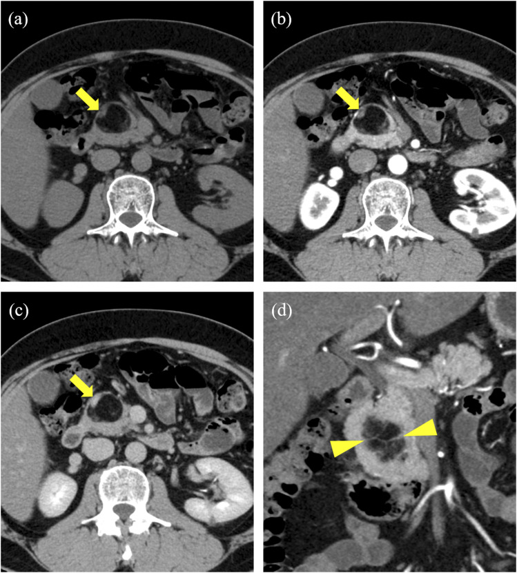
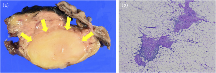
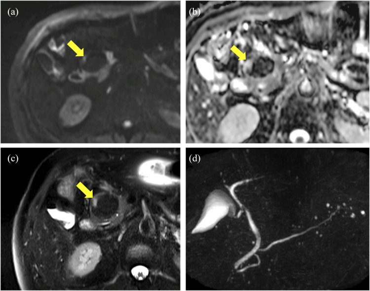
Lipomatous pseudohypertrophy of the pancreas (LPH) is a rare disease in which the pancreatic parenchyma is replaced with mature adipose tissue. It is an idiopathic condition whose diagnosis is made based on histopathological analyses. Herein, we report the case of a 50-year-old male patient with a lipomatous mass in the head of the pancreas on computed tomography for close examination of a renal tumor. We suspected liposarcoma, and laparotomy was performed. However, histological analyses revealed LPH. Several imaging findings of LPH can enable a noninvasive diagnosis and help its clinical approach.
模仿脂肪肉瘤的胰腺脂肪瘤性假肥大:病例报告。
胰腺脂肪瘤性假性肥厚(LPH)是一种罕见疾病,在这种疾病中,胰腺实质被成熟的脂肪组织取代。这是一种特发性疾病,其诊断依据是组织病理学分析。在此,我们报告了一例 50 岁男性患者的病例,他在计算机断层扫描中发现胰腺头部有脂肪瘤肿块,仔细检查后发现是肾肿瘤。我们怀疑是脂肪肉瘤,于是进行了开腹手术。然而,组织学分析却显示为 LPH。LPH的几种成像结果可以实现无创诊断,并有助于临床治疗。
本文章由计算机程序翻译,如有差异,请以英文原文为准。
求助全文
约1分钟内获得全文
求助全文

 求助内容:
求助内容: 应助结果提醒方式:


