{"title":"First tarsometatarsal joint mobility in hallux valgus during gait: A synchronized ultrasound and three-dimensional motion capture analysis","authors":"Tsubasa Tashiro, Yasunari Ikuta, Noriaki Maeda, Satoshi Arima, Masanori Morikawa, Kazuki Kaneda, Honoka Ishihara, Shogo Tsutsumi, Miki Kawai, Andreas Brand, Tomoyuki Nakasa, Nobuo Adachi, Makoto Komiya, Yukio Urabe","doi":"10.1007/s10396-024-01414-2","DOIUrl":null,"url":null,"abstract":"<h3 data-test=\"abstract-sub-heading\">Purpose</h3><p>To quantify the vertical translation between the first metatarsal and medial cuneiform during the stance phase of gait in young individuals with and without hallux valgus.</p><h3 data-test=\"abstract-sub-heading\">Design</h3><p>This cross-sectional observational study included 34 young adults (male, <i>n</i> = 4; female, <i>n</i> = 30) who were divided into three groups according to the hallux valgus angle: control (< 20°, <i>n</i> = 13), mild hallux valgus (≥ 20° to < 30°, <i>n</i> = 12), and moderate hallux valgus (≥ 30°, <i>n</i> = 9). The mobility of the first tarsometatarsal joint was evaluated during the stance phase using B-mode ultrasound synchronized with a motion analysis system.</p><h3 data-test=\"abstract-sub-heading\">Results</h3><p>The medial cuneiform shifted more plantar during the early phase in mild hallux valgus and during the middle and terminal phases in moderate hallux valgus than in control. The severity of the hallux valgus was correlated with a trend toward plantar shift of the medial cuneiform. The first metatarsal was located more dorsal than the medial cuneiform; however, there was no significant variation. No significant differences in the peak ankle plantarflexion angle and moment were noted between the groups.</p><h3 data-test=\"abstract-sub-heading\">Conclusion</h3><p>The hypermobility of the first tarsometatarsal joint, especially plantar displacement of the medial cuneiform in the sagittal plane, was found in young individuals with hallux valgus during the stance phase of gait, and the mobility increased with the severity of hallux valgus. Our findings suggest the significance of preventing hallux valgus deformity early in life.</p>","PeriodicalId":50130,"journal":{"name":"Journal of Medical Ultrasonics","volume":null,"pages":null},"PeriodicalIF":1.9000,"publicationDate":"2024-03-28","publicationTypes":"Journal Article","fieldsOfStudy":null,"isOpenAccess":false,"openAccessPdf":"","citationCount":"0","resultStr":null,"platform":"Semanticscholar","paperid":null,"PeriodicalName":"Journal of Medical Ultrasonics","FirstCategoryId":"3","ListUrlMain":"https://doi.org/10.1007/s10396-024-01414-2","RegionNum":4,"RegionCategory":"医学","ArticlePicture":[],"TitleCN":null,"AbstractTextCN":null,"PMCID":null,"EPubDate":"","PubModel":"","JCR":"Q3","JCRName":"RADIOLOGY, NUCLEAR MEDICINE & MEDICAL IMAGING","Score":null,"Total":0}
引用次数: 0
Abstract
Purpose
To quantify the vertical translation between the first metatarsal and medial cuneiform during the stance phase of gait in young individuals with and without hallux valgus.
Design
This cross-sectional observational study included 34 young adults (male, n = 4; female, n = 30) who were divided into three groups according to the hallux valgus angle: control (< 20°, n = 13), mild hallux valgus (≥ 20° to < 30°, n = 12), and moderate hallux valgus (≥ 30°, n = 9). The mobility of the first tarsometatarsal joint was evaluated during the stance phase using B-mode ultrasound synchronized with a motion analysis system.
Results
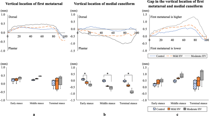
The medial cuneiform shifted more plantar during the early phase in mild hallux valgus and during the middle and terminal phases in moderate hallux valgus than in control. The severity of the hallux valgus was correlated with a trend toward plantar shift of the medial cuneiform. The first metatarsal was located more dorsal than the medial cuneiform; however, there was no significant variation. No significant differences in the peak ankle plantarflexion angle and moment were noted between the groups.
Conclusion
The hypermobility of the first tarsometatarsal joint, especially plantar displacement of the medial cuneiform in the sagittal plane, was found in young individuals with hallux valgus during the stance phase of gait, and the mobility increased with the severity of hallux valgus. Our findings suggest the significance of preventing hallux valgus deformity early in life.
期刊介绍:
The Journal of Medical Ultrasonics is the official journal of the Japan Society of Ultrasonics in Medicine. The main purpose of the journal is to provide forum for the publication of papers documenting recent advances and new developments in the entire field of ultrasound in medicine and biology, encompassing both the medical and the engineering aspects of the science.The journal welcomes original articles, review articles, images, and letters to the editor.The journal also provides state-of-the-art information such as announcements from the boards and the committees of the society.
 求助内容:
求助内容: 应助结果提醒方式:


