Thomas J M Verlinden, Wouter H Lamers, Andreas Herrler, S Eleonore Köhler
{"title":"The differences in the anatomy of the thoracolumbar and sacral autonomic outflow are quantitative.","authors":"Thomas J M Verlinden, Wouter H Lamers, Andreas Herrler, S Eleonore Köhler","doi":"10.1007/s10286-024-01023-6","DOIUrl":null,"url":null,"abstract":"<p><strong>Purpose: </strong>We have re-evaluated the anatomical arguments that underlie the division of the spinal visceral outflow into sympathetic and parasympathetic divisions.</p><p><strong>Methodology: </strong>Using a systematic literature search, we mapped the location of catecholaminergic neurons throughout the mammalian peripheral nervous system. Subsequently, a narrative method was employed to characterize segment-dependent differences in the location of preganglionic cell bodies and the composition of white and gray rami communicantes.</p><p><strong>Results and conclusion: </strong>One hundred seventy studies were included in the systematic review, providing information on 389 anatomical structures. Catecholaminergic nerve fibers are present in most spinal and all cranial nerves and ganglia, including those that are known for their parasympathetic function. Along the entire spinal autonomic outflow pathways, proximal and distal catecholaminergic cell bodies are common in the head, thoracic, and abdominal and pelvic region, which invalidates the \"short-versus-long preganglionic neuron\" argument. Contrary to the classically confined outflow levels T1-L2 and S2-S4, preganglionic neurons have been found in the resulting lumbar gap. Preganglionic cell bodies that are located in the intermediolateral zone of the thoracolumbar spinal cord gradually nest more ventrally within the ventral motor nuclei at the lumbar and sacral levels, and their fibers bypass the white ramus communicans and sympathetic trunk to emerge directly from the spinal roots. Bypassing the sympathetic trunk, therefore, is not exclusive for the sacral outflow. We conclude that the autonomic outflow displays a conserved architecture along the entire spinal axis, and that the perceived differences in the anatomy of the autonomic thoracolumbar and sacral outflow are quantitative.</p>","PeriodicalId":10168,"journal":{"name":"Clinical Autonomic Research","volume":" ","pages":"79-97"},"PeriodicalIF":3.4000,"publicationDate":"2024-02-01","publicationTypes":"Journal Article","fieldsOfStudy":null,"isOpenAccess":false,"openAccessPdf":"https://www.ncbi.nlm.nih.gov/pmc/articles/PMC10944453/pdf/","citationCount":"0","resultStr":null,"platform":"Semanticscholar","paperid":null,"PeriodicalName":"Clinical Autonomic Research","FirstCategoryId":"3","ListUrlMain":"https://doi.org/10.1007/s10286-024-01023-6","RegionNum":3,"RegionCategory":"医学","ArticlePicture":[],"TitleCN":null,"AbstractTextCN":null,"PMCID":null,"EPubDate":"2024/2/25 0:00:00","PubModel":"Epub","JCR":"Q1","JCRName":"CLINICAL NEUROLOGY","Score":null,"Total":0}
引用次数: 0
Abstract
Purpose: We have re-evaluated the anatomical arguments that underlie the division of the spinal visceral outflow into sympathetic and parasympathetic divisions.
Methodology: Using a systematic literature search, we mapped the location of catecholaminergic neurons throughout the mammalian peripheral nervous system. Subsequently, a narrative method was employed to characterize segment-dependent differences in the location of preganglionic cell bodies and the composition of white and gray rami communicantes.
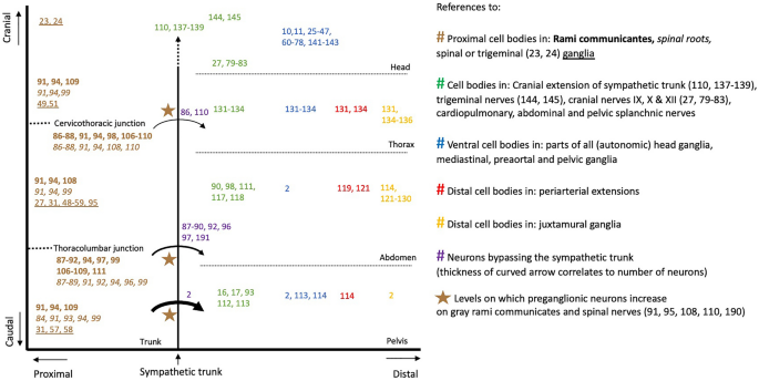
Results and conclusion: One hundred seventy studies were included in the systematic review, providing information on 389 anatomical structures. Catecholaminergic nerve fibers are present in most spinal and all cranial nerves and ganglia, including those that are known for their parasympathetic function. Along the entire spinal autonomic outflow pathways, proximal and distal catecholaminergic cell bodies are common in the head, thoracic, and abdominal and pelvic region, which invalidates the "short-versus-long preganglionic neuron" argument. Contrary to the classically confined outflow levels T1-L2 and S2-S4, preganglionic neurons have been found in the resulting lumbar gap. Preganglionic cell bodies that are located in the intermediolateral zone of the thoracolumbar spinal cord gradually nest more ventrally within the ventral motor nuclei at the lumbar and sacral levels, and their fibers bypass the white ramus communicans and sympathetic trunk to emerge directly from the spinal roots. Bypassing the sympathetic trunk, therefore, is not exclusive for the sacral outflow. We conclude that the autonomic outflow displays a conserved architecture along the entire spinal axis, and that the perceived differences in the anatomy of the autonomic thoracolumbar and sacral outflow are quantitative.
期刊介绍:
Clinical Autonomic Research aims to draw together and disseminate research work from various disciplines and specialties dealing with clinical problems resulting from autonomic dysfunction. Areas to be covered include: cardiovascular system, neurology, diabetes, endocrinology, urology, pain disorders, ophthalmology, gastroenterology, toxicology and clinical pharmacology, skin infectious diseases, renal disease.
This journal is an essential source of new information for everyone working in areas involving the autonomic nervous system. A major feature of Clinical Autonomic Research is its speed of publication coupled with the highest refereeing standards.
 求助内容:
求助内容: 应助结果提醒方式:


