{"title":"Diagnostic value of high-frequency ultrasound in omohyoid muscle syndrome","authors":"Liyuan Cui, Ling Wang, Tiezheng Wang, Yeting Wang, Wen Chen, Hengtao Qi","doi":"10.1007/s10396-023-01407-7","DOIUrl":null,"url":null,"abstract":"<h3 data-test=\"abstract-sub-heading\">Purpose</h3><p>To investigate the diagnostic value of high-frequency ultrasound in omohyoid muscle syndrome.</p><h3 data-test=\"abstract-sub-heading\">Material and methods</h3><p>A retrospective analysis of 11 patients diagnosed with omohyoid muscle syndrome was carried out, and the characteristics of high-frequency ultrasound images were summarized.</p><h3 data-test=\"abstract-sub-heading\">Results</h3><p>Ultrasonography of the omohyoid muscle showed a narrow band of hypoechoic muscle bundle. The ultrasonographic manifestation of omohyoid muscle syndrome showed a thickening of the omohyoid muscle on the affected side. The omohyoid muscle on the affected side bulged forward during swallowing and lifted the overlying sternocleidomastoid muscle. The difference between the thickness of the omohyoid muscle intermediate tendon on the affected side and the healthy side at rest was statistically significant (<i>t</i> = 58.23, <i>P</i> < 0.001). The difference between the thickness of the affected omohyoid muscle intermediate tendon at rest and during swallowing was statistically significant (<i>t</i> = 14.57, <i>P</i> < 0.001). There was no statistically significant difference between the thickness of the omohyoid muscle intermediate tendon on the healthy side at rest and during swallowing (<i>t</i> = 0.56, <i>P</i> > 0.05).</p><h3 data-test=\"abstract-sub-heading\">Conclusion</h3><p>High-frequency ultrasound is the preferred imaging method in the diagnosis of omohyoid muscle syndrome.</p>","PeriodicalId":50130,"journal":{"name":"Journal of Medical Ultrasonics","volume":null,"pages":null},"PeriodicalIF":1.9000,"publicationDate":"2024-01-31","publicationTypes":"Journal Article","fieldsOfStudy":null,"isOpenAccess":false,"openAccessPdf":"","citationCount":"0","resultStr":null,"platform":"Semanticscholar","paperid":null,"PeriodicalName":"Journal of Medical Ultrasonics","FirstCategoryId":"3","ListUrlMain":"https://doi.org/10.1007/s10396-023-01407-7","RegionNum":4,"RegionCategory":"医学","ArticlePicture":[],"TitleCN":null,"AbstractTextCN":null,"PMCID":null,"EPubDate":"","PubModel":"","JCR":"Q3","JCRName":"RADIOLOGY, NUCLEAR MEDICINE & MEDICAL IMAGING","Score":null,"Total":0}
引用次数: 0
Abstract
Purpose
To investigate the diagnostic value of high-frequency ultrasound in omohyoid muscle syndrome.
Material and methods
A retrospective analysis of 11 patients diagnosed with omohyoid muscle syndrome was carried out, and the characteristics of high-frequency ultrasound images were summarized.
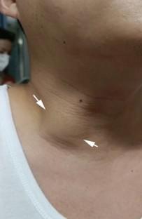
Results
Ultrasonography of the omohyoid muscle showed a narrow band of hypoechoic muscle bundle. The ultrasonographic manifestation of omohyoid muscle syndrome showed a thickening of the omohyoid muscle on the affected side. The omohyoid muscle on the affected side bulged forward during swallowing and lifted the overlying sternocleidomastoid muscle. The difference between the thickness of the omohyoid muscle intermediate tendon on the affected side and the healthy side at rest was statistically significant (t = 58.23, P < 0.001). The difference between the thickness of the affected omohyoid muscle intermediate tendon at rest and during swallowing was statistically significant (t = 14.57, P < 0.001). There was no statistically significant difference between the thickness of the omohyoid muscle intermediate tendon on the healthy side at rest and during swallowing (t = 0.56, P > 0.05).
Conclusion
High-frequency ultrasound is the preferred imaging method in the diagnosis of omohyoid muscle syndrome.
期刊介绍:
The Journal of Medical Ultrasonics is the official journal of the Japan Society of Ultrasonics in Medicine. The main purpose of the journal is to provide forum for the publication of papers documenting recent advances and new developments in the entire field of ultrasound in medicine and biology, encompassing both the medical and the engineering aspects of the science.The journal welcomes original articles, review articles, images, and letters to the editor.The journal also provides state-of-the-art information such as announcements from the boards and the committees of the society.
 求助内容:
求助内容: 应助结果提醒方式:


