{"title":"Case report: Urothelial carcinoma of the renal pelvis with trophoblastic differentiation: A rare case report and review of literature.","authors":"Zhuo Wang, Jinsui Wang, Wenwen Zhang, Daoying Wang, Xiaojun Wang, Xiaoqin Liang","doi":"10.3389/pore.2023.1610856","DOIUrl":null,"url":null,"abstract":"<p><p>We report a rare case of urothelial carcinoma (UC) of the renal pelvis with trophoblastic differentiation that occurred in a 55-year-old male patient. The patient presented with gross hematuria and paroxysmal lumbago pain 5 months ago. The enhanced computed tomography (CT) scan demonstrated a large space occupying lesion in the left kidney and multiple retroperitoneal lymph node enlargements. Histologically, high-grade infiltrating urothelial carcinoma (HGUC) contained giant cells which were positive for beta-human chorionic gonadotropin (β-hCG). Three weeks after resection, positron emission tomography and computed tomography (PET-CT) scan showed multiple nodules of metastasis in the left renal region, extensive systemic muscle, bone, lymph node, liver and bilateral lung metastases. The patient underwent bladder perfusion chemotherapy and gemcitabine combined with cisplatin chemotherapy regimens. This is the eighth documented case of UC of the renal pelvis with trophoblastic differentiation. Due to its rarity and extremely poor prognosis, it is important to clarify the characteristics of the disease and make an accurate and prompt diagnosis.</p>","PeriodicalId":19981,"journal":{"name":"Pathology & Oncology Research","volume":"29 ","pages":"1610856"},"PeriodicalIF":2.3000,"publicationDate":"2023-01-01","publicationTypes":"Journal Article","fieldsOfStudy":null,"isOpenAccess":false,"openAccessPdf":"https://www.ncbi.nlm.nih.gov/pmc/articles/PMC9950125/pdf/","citationCount":"0","resultStr":null,"platform":"Semanticscholar","paperid":null,"PeriodicalName":"Pathology & Oncology Research","FirstCategoryId":"3","ListUrlMain":"https://doi.org/10.3389/pore.2023.1610856","RegionNum":4,"RegionCategory":"医学","ArticlePicture":[],"TitleCN":null,"AbstractTextCN":null,"PMCID":null,"EPubDate":"","PubModel":"","JCR":"Q3","JCRName":"ONCOLOGY","Score":null,"Total":0}
引用次数: 0
Abstract
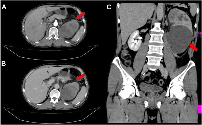
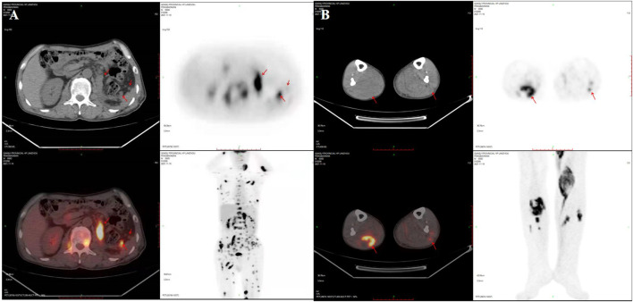
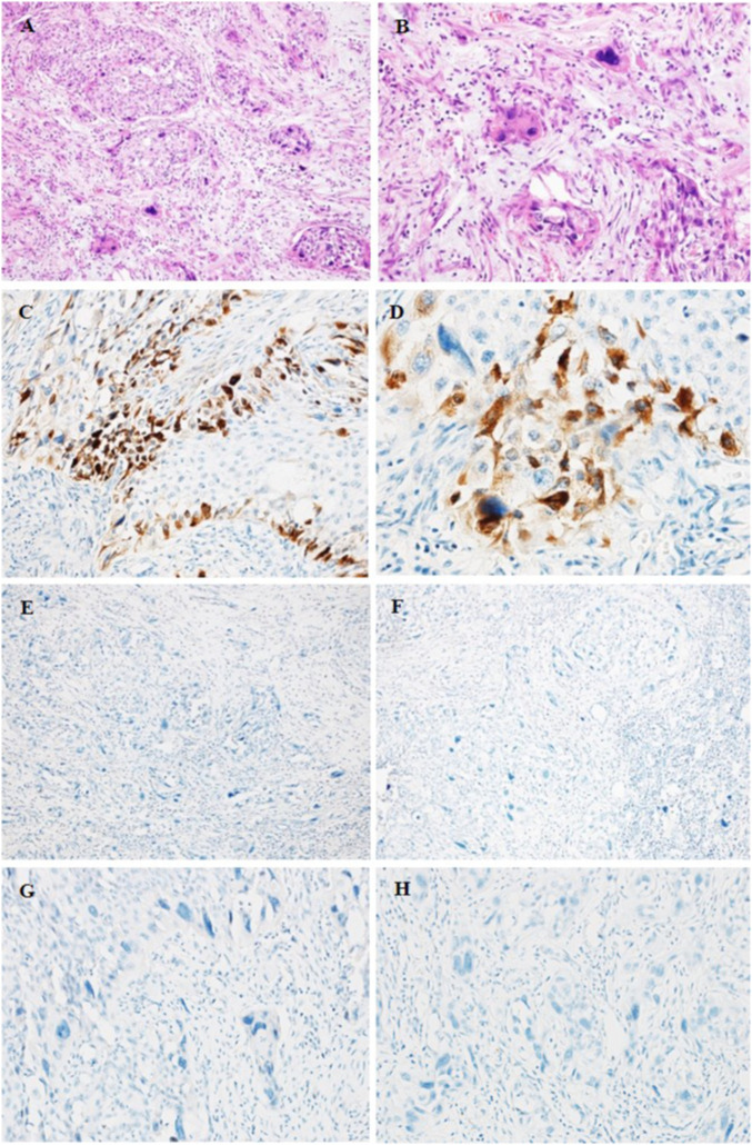
We report a rare case of urothelial carcinoma (UC) of the renal pelvis with trophoblastic differentiation that occurred in a 55-year-old male patient. The patient presented with gross hematuria and paroxysmal lumbago pain 5 months ago. The enhanced computed tomography (CT) scan demonstrated a large space occupying lesion in the left kidney and multiple retroperitoneal lymph node enlargements. Histologically, high-grade infiltrating urothelial carcinoma (HGUC) contained giant cells which were positive for beta-human chorionic gonadotropin (β-hCG). Three weeks after resection, positron emission tomography and computed tomography (PET-CT) scan showed multiple nodules of metastasis in the left renal region, extensive systemic muscle, bone, lymph node, liver and bilateral lung metastases. The patient underwent bladder perfusion chemotherapy and gemcitabine combined with cisplatin chemotherapy regimens. This is the eighth documented case of UC of the renal pelvis with trophoblastic differentiation. Due to its rarity and extremely poor prognosis, it is important to clarify the characteristics of the disease and make an accurate and prompt diagnosis.
期刊介绍:
Pathology & Oncology Research (POR) is an interdisciplinary Journal at the interface of pathology and oncology including the preclinical and translational research, diagnostics and therapy. Furthermore, POR is an international forum for the rapid communication of reviews, original research, critical and topical reports with excellence and novelty. Published quarterly, POR is dedicated to keeping scientists informed of developments on the selected biomedical fields bridging the gap between basic research and clinical medicine. It is a special aim for POR to promote pathological and oncological publishing activity of colleagues in the Central and East European region. The journal will be of interest to pathologists, and a broad range of experimental and clinical oncologists, and related experts. POR is supported by an acknowledged international advisory board and the Arányi Fundation for modern pathology.
 求助内容:
求助内容: 应助结果提醒方式:


