Imaging features, clinical characteristics and neonatal outcomes of pregnancy luteoma: A case series and literature review

IF 3.5 2区 医学 Q1 OBSTETRICS & GYNECOLOGY
Jian-Hong Shang, Cai-Xin Huang, Qiao Zheng, Jie-Ling Feng, Ke He, Hong-Ning Xie
{"title":"Imaging features, clinical characteristics and neonatal outcomes of pregnancy luteoma: A case series and literature review","authors":"Jian-Hong Shang,&nbsp;Cai-Xin Huang,&nbsp;Qiao Zheng,&nbsp;Jie-Ling Feng,&nbsp;Ke He,&nbsp;Hong-Ning Xie","doi":"10.1111/aogs.14672","DOIUrl":null,"url":null,"abstract":"<div>\n \n \n <section>\n \n <h3> Introduction</h3>\n \n <p>This study aimed to investigate the imaging features, clinical characteristics and neonatal outcomes of pregnancy luteoma.</p>\n </section>\n \n <section>\n \n <h3> Material and methods</h3>\n \n <p>We retrospectively analyzed patients with pregnancy luteoma admitted to the First Affiliated Hospital of Sun Yat-sen University between January 2003 and December 2022. We recorded their imaging features, clinical characteristics and neonatal outcomes. Additionally, we reviewed relevant studies in the field.</p>\n </section>\n \n <section>\n \n <h3> Results</h3>\n \n <p>In total, 127 cases were identified, including eight from our hospital and 119 from the literature. Most patients (93/127, 73.23%) were of reproductive age, 20–40 years old, and 66% were parous. Maternal hirsutism or virilization (such as deepening voice, acne, facial hair growth and clitoromegaly) was observed in 29.92% (38/127), whereas 59.06% of patients (75/127) were asymptomatic. Abdominal pain was reported in 13 patients due to compression, torsion or combined ectopic pregnancy. The pregnancy luteomas, primarily discovered during the third trimester (79/106, 74.53%), varied in size ranging from 10 mm to 20 cm in diameter. Seventy-five cases were incidentally detected during cesarean section or postpartum tubal ligation, and 39 were identified through imaging or physical examination during pregnancy. Approximately 26.61% of patients had bilateral lesions. The majority of pregnancy luteomas were solid and well-defined (94/107, 87.85%), with 43.06% (31/72) displaying multiple solid and well-circumscribed nodules. Elevated serum androgen levels (reaching values between 1.24 and 1529 times greater than normal values for term gestation) were observed in patients with hirsutism or virilization, with a larger lesion diameter (<i>P</i> &lt; 0.001) and a higher prevalence of bilateral lesions (<i>P</i> &lt; 0.001). Among the female infants born to masculinized mothers, 68.18% (15/22) were virilized. Information of imaging features was complete in 22 cases. Ultrasonography revealed well-demarcated hypoechoic solid masses with rich blood supply in 12 of 19 cases (63.16%). Nine patients underwent magnetic resonance imaging (MRI) or computed tomography (CT), and six exhibited solid masses, including three with multi-nodular solid masses.</p>\n </section>\n \n <section>\n \n <h3> Conclusions</h3>\n \n <p>Pregnancy luteomas mainly manifest as well-defined, hypoechoic and hypervascular solid masses. MRI and CT are superior to ultrasonography in displaying the imaging features of multiple nodules. Maternal masculinization and solid masses with multiple nodules on imaging may help diagnose this rare disease.</p>\n </section>\n </div>","PeriodicalId":6990,"journal":{"name":"Acta Obstetricia et Gynecologica Scandinavica","volume":"103 4","pages":"740-750"},"PeriodicalIF":3.5000,"publicationDate":"2023-09-14","publicationTypes":"Journal Article","fieldsOfStudy":null,"isOpenAccess":false,"openAccessPdf":"https://onlinelibrary.wiley.com/doi/epdf/10.1111/aogs.14672","citationCount":"0","resultStr":null,"platform":"Semanticscholar","paperid":null,"PeriodicalName":"Acta Obstetricia et Gynecologica Scandinavica","FirstCategoryId":"3","ListUrlMain":"https://onlinelibrary.wiley.com/doi/10.1111/aogs.14672","RegionNum":2,"RegionCategory":"医学","ArticlePicture":[],"TitleCN":null,"AbstractTextCN":null,"PMCID":null,"EPubDate":"","PubModel":"","JCR":"Q1","JCRName":"OBSTETRICS & GYNECOLOGY","Score":null,"Total":0}
引用次数: 0

Abstract

Introduction

This study aimed to investigate the imaging features, clinical characteristics and neonatal outcomes of pregnancy luteoma.

Material and methods

We retrospectively analyzed patients with pregnancy luteoma admitted to the First Affiliated Hospital of Sun Yat-sen University between January 2003 and December 2022. We recorded their imaging features, clinical characteristics and neonatal outcomes. Additionally, we reviewed relevant studies in the field.

Results

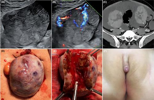
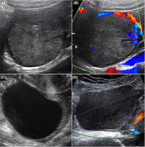
In total, 127 cases were identified, including eight from our hospital and 119 from the literature. Most patients (93/127, 73.23%) were of reproductive age, 20–40 years old, and 66% were parous. Maternal hirsutism or virilization (such as deepening voice, acne, facial hair growth and clitoromegaly) was observed in 29.92% (38/127), whereas 59.06% of patients (75/127) were asymptomatic. Abdominal pain was reported in 13 patients due to compression, torsion or combined ectopic pregnancy. The pregnancy luteomas, primarily discovered during the third trimester (79/106, 74.53%), varied in size ranging from 10 mm to 20 cm in diameter. Seventy-five cases were incidentally detected during cesarean section or postpartum tubal ligation, and 39 were identified through imaging or physical examination during pregnancy. Approximately 26.61% of patients had bilateral lesions. The majority of pregnancy luteomas were solid and well-defined (94/107, 87.85%), with 43.06% (31/72) displaying multiple solid and well-circumscribed nodules. Elevated serum androgen levels (reaching values between 1.24 and 1529 times greater than normal values for term gestation) were observed in patients with hirsutism or virilization, with a larger lesion diameter (P < 0.001) and a higher prevalence of bilateral lesions (P < 0.001). Among the female infants born to masculinized mothers, 68.18% (15/22) were virilized. Information of imaging features was complete in 22 cases. Ultrasonography revealed well-demarcated hypoechoic solid masses with rich blood supply in 12 of 19 cases (63.16%). Nine patients underwent magnetic resonance imaging (MRI) or computed tomography (CT), and six exhibited solid masses, including three with multi-nodular solid masses.

Conclusions

Pregnancy luteomas mainly manifest as well-defined, hypoechoic and hypervascular solid masses. MRI and CT are superior to ultrasonography in displaying the imaging features of multiple nodules. Maternal masculinization and solid masses with multiple nodules on imaging may help diagnose this rare disease.
Abstract Image

Abstract Image

妊娠黄体瘤的影像学特征、临床特点和新生儿预后:病例系列和文献综述
引言 本研究旨在探讨妊娠黄体瘤的影像学特征、临床特点和新生儿预后。 材料与方法 我们回顾性分析了2003年1月至2022年12月期间中山大学附属第一医院收治的妊娠黄体瘤患者。我们记录了他们的影像学特征、临床特点和新生儿结局。此外,我们还回顾了该领域的相关研究。 结果 共发现 127 例病例,其中 8 例来自本院,119 例来自文献。大多数患者(93/127,73.23%)处于育龄期,年龄在 20-40 岁之间,66% 为奇数。29.92%的患者(38/127)观察到母体多毛或男性化(如声音加深、痤疮、面部毛发生长和阴蒂肥大),而59.06%的患者(75/127)无症状。13例患者因压迫、扭转或合并异位妊娠而出现腹痛。妊娠黄体瘤主要是在怀孕三个月时发现的(79/106,74.53%),大小不一,直径从 10 毫米到 20 厘米不等。75例是在剖腹产或产后输卵管结扎时偶然发现的,39例是在怀孕期间通过造影或体检发现的。约 26.61% 的患者为双侧病变。大多数妊娠黄体瘤为实性,轮廓清晰(94/107,87.85%),43.06%(31/72)的黄体瘤为多发性实性环状结节。多毛症或男性化患者的血清雄激素水平升高(达到足月妊娠正常值的 1.24 至 1529 倍),病变直径更大(P <;0.001),双侧病变发生率更高(P <;0.001)。在母亲男性化的女婴中,68.18%(15/22)为男性化。22 例病例的影像学特征信息完整。超声波检查显示,19 例中有 12 例(63.16%)的实性肿块边界清晰,血供丰富。9 例患者接受了磁共振成像(MRI)或计算机断层扫描(CT),其中 6 例显示实性肿块,包括 3 例多结节实性肿块。 结论 妊娠黄体瘤主要表现为界限清楚、低回声和高血管实性肿块。磁共振成像和 CT 在显示多结节的影像特征方面优于超声波检查。母体男性化和实性肿块伴多发结节的影像学表现有助于诊断这种罕见疾病。
本文章由计算机程序翻译,如有差异,请以英文原文为准。
求助全文
约1分钟内获得全文 求助全文
来源期刊
CiteScore
8.00
自引率
4.70%
发文量
180
审稿时长
3-6 weeks
期刊介绍: Published monthly, Acta Obstetricia et Gynecologica Scandinavica is an international journal dedicated to providing the very latest information on the results of both clinical, basic and translational research work related to all aspects of women’s health from around the globe. The journal regularly publishes commentaries, reviews, and original articles on a wide variety of topics including: gynecology, pregnancy, birth, female urology, gynecologic oncology, fertility and reproductive biology.
×
引用
GB/T 7714-2015
复制
MLA
复制
APA
复制
导出至
BibTeX EndNote RefMan NoteFirst NoteExpress
×
提示
您的信息不完整,为了账户安全,请先补充。
现在去补充
×
提示
您因"违规操作"
具体请查看互助需知
我知道了
×
提示
确定
请完成安全验证×
copy
已复制链接
快去分享给好友吧!
我知道了
右上角分享
点击右上角分享
0
联系我们:info@booksci.cn Book学术提供免费学术资源搜索服务,方便国内外学者检索中英文文献。致力于提供最便捷和优质的服务体验。 Copyright © 2023 布克学术 All rights reserved.
京ICP备2023020795号-1
ghs 京公网安备 11010802042870号
Book学术文献互助
Book学术文献互助群
群 号:481959085
Book学术官方微信